Steerable extendable devices

a technology of extendable devices and steering devices, which is applied in the field of medical devices, robotics, oil and gas, civil engineering, disaster robotics, etc., can solve the problems of multiple punctures or incisions, time-consuming, and inability to access the target region of current devices, so as to avoid iterative, time-consuming, and potentially traumatic manipulation

Inactive Publication Date: 2021-09-23
SOUTHERN METHODIST UNIVERSITY
View PDF0 Cites 1 Cited by
  • Summary
  • Abstract
  • Description
  • Claims
  • Application Information

AI Technical Summary

Benefits of technology

[0008]According to some embodiments of the invention, a stable, controllable cannula is provided that follows an optimal 3-D path—through gas or liquid-filled volumes or solid tissue—to reach virtually any target at any approach angle, and do so without iterative, time-consuming, and potentially traumatic manipulation by the clinician. FIG. 4(a-c) depicts this ideal sequence, in which the device extends along a path while steered and assembled robotically in-vivo so as to grow into a complex, often curvilinear 3-D shape within the body while the more distal regions of the device retain their original shape.
[0009]Such a device, which we term a “distally-assembled steerable cannula” (hereinafter, “DASC”), is fundamentally different from existing steerable needles, cannulas, catheters, and snake robots that rely on distally sliding and articulating. DASC is unique in several aspects. For example, it may be deployed within a patient's body entirely through distal growth, extending at its distal tip to follow a controlled 3-D path, while shape is maintained everywhere along the device. Moreover, it may be assembled in vivo from multiple, discrete pieces (interlocking segments or rings), be continuously assembled from a strip, or grow as an everting, steerable tube, offering an unprecedented number of possible 3-D shapes. These shapes may include multiple bends in multiple planes at various locations along the length of the cannula. Moreover, the cannula can vary its overall length up to a maximum. These attributes together can greatly facilitate operation within confined spaces. DASC provides an enormous number of degrees of freedom, yet does not require many actuators, since the mechanisms providing those degrees of freedom are self-locking and are accessed sequentially. DASC offers the ability to make tight turns (e.g., a 2:1 radius of curvature to O.D. ratio); for example, a 0.120″ O.D. device could make a full 180° turn in a space only ˜0.7″ wide. DASC also provides an unusually large lumen / working channel (e.g., a 0.9:1.0 lumen / O.D. ratio)—comparable to a non-steerable catheter—enabling more instruments to be used simultaneously; improving endoscopic visualization, irrigation, and aspiration; and allowing more tissue to be excised (e.g., for biopsy or tumor resection).
[0010]DASC can serve as a stable, passive conduit that provides access and support to other devices (e.g., articulated endoscopes, forceps, bipolar diathermy and monopolar cautery devices, laser fibers, graspers, dissectors, scissors, knives, needles, needle drivers, spatulas, or other instruments, at least one of which can be used at a time, and which can be rapidly exchanged), for example, in endoscopic surgery, laparoscopic, single port, and natural orifice translumenal endoscopic surgery, or to infuse or aspirate liquids. However, unlike prior-art passive devices, DASC can be easily re-shaped distally without having to first withdraw it and re-insert it during a procedure. Thus, DASC can be disassembled partially and then reassembled such that the distal end moves to a new position and / or changes its approach angle to the target region, providing, for example, a stable platform for procedures which cover multiple sites or wide areas.
[0019]In another embodiment, the present invention includes a method of making an elongatable, steerable cannula capable of growing from a proximal end to a distal end through a process of assembly wherein one or more segments are added to the distal end of the cannula, the method comprising: obtaining a first flexible segment substantially cylindrical in axial cross section when uncompressed and substantially elliptical when compressed, having a first lumen therethrough, the first segment having a first proximal face substantially perpendicular to a first substantially cylindrical axis and provided with one or more first male coupling elements, and a first distal face substantially non-perpendicular to the substantially cylindrical axis and provided with a first plurality of female coupling elements; positioning a second flexible segment substantially cylindrical in axial cross section when uncompressed and substantially elliptical when compressed, having a second lumen therethrough, the second segment having a second proximal face substantially non-perpendicular to a second substantially cylindrical axis and provided with one or more second male coupling elements, and a second distal face substantially perpendicular to the second substantially cylindrical axis and provided with a second plurality of female coupling elements; inserting a flexible shaft provided with grippers able to grip said first and said second segment to compress and allow to decompress said segments, swivel or allow to swivel said segments about axes substantially parallel to the minor axes of said first and second segments, and transport said segments through said lumen; wherein said flexible shaft transports said second segment through said lumen of said first segment, rotates said second segment as required around an axis substantially coincident with the longitudinal axis of the flexible shaft, brings the proximal face of said second segment in contact with the distal face of said first segment, then allows said second segment to decompress while allowing two of said second male coupling elements to enter two of said first plurality of female coupling elements from the inside and two of said second male coupling elements to enter two of said first plurality of female coupling elements from the outside; and wherein said flexible shaft transports said first segment through said lumen of said second segment, rotates said first segment as required around an axis substantially coincident with the longitudinal axis of the flexible shaft, brings the proximal face of said first segment in contact with the distal face of said second segment, then allows said first segment to decompress while allowing two of said first male coupling elements to enter two of said second plurality of female coupling elements from the inside and two of said first male coupling elements to enter two of said second plurality of female coupling elements from the outside.

Problems solved by technology

Yet due to obstructions such as bone or sensitive organs, current devices may be unable to access a target region, or only do so sub-optimally.
A more invasive method, a riskier approach, or a worse outcome is thus sometimes unavoidable.
Moreover, in a number of procedures for which multiple targeted regions within the body need to be accessed, it can be time-consuming and involve multiple punctures or incisions, even if access to a single region would be straightforward.
These issues arise from the fact that many instruments are substantially rigid and straight and follow substantially straight paths within the body, whether within a hollow (i.e., gas- or liquid-filled) organ or lumen, or in solid tissue.
Moreover, even instruments known to the art that are not rigid and / or not straight can also have difficulty in accessing certain regions of the body, as the following examples will illustrate.
To date such devices have been problematic and many remain experimental.
Spinning steerable needles with asymmetric tips [1,2,3,4] offer small gauge sizes but have very large curvature / outer diameter (O.D.) ratios (e.g., 70:1 [5]), can only be deployed within solid tissue, and are difficult to control accurately due to varying tissue properties [6], etc.

Method used


Image

  • Steerable extendable devices
  • Steerable extendable devices
  • Steerable extendable devices

Examples


1st embodiment

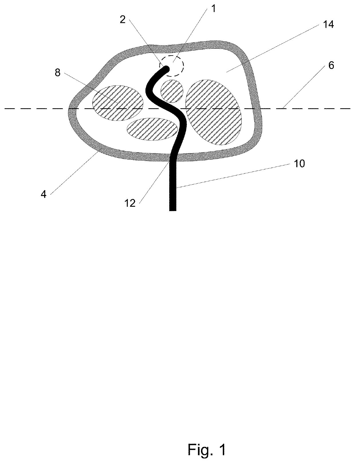
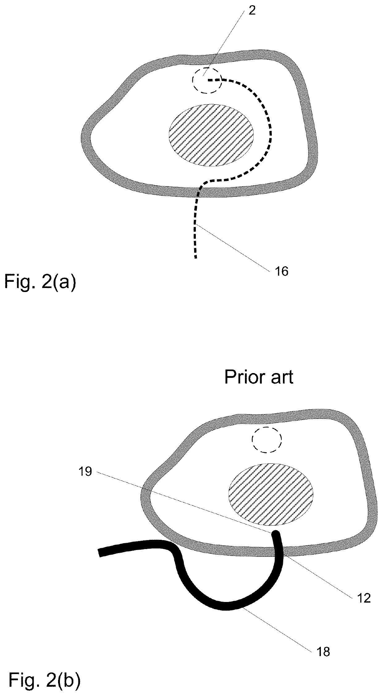
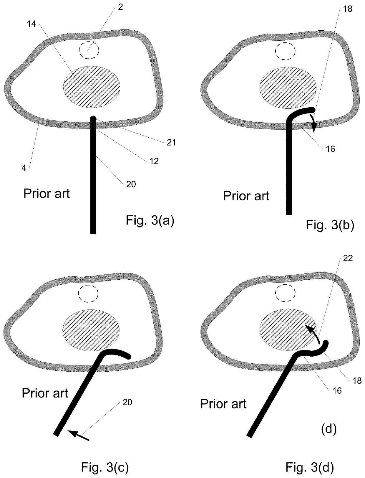
[0120]In a 1st embodiment of the invention, an instrument enters the body and is delivered along a desired path such as path 16 of FIG. 2(a) as illustrated in FIGS. 4(a-c) to reach region 2. The path may be determined, for example, using imaging such as CT or MM. In FIG. 4(a), instrument 30 enters the skin 4 through puncture site 12 and begins to follow path 16, curving as it grows or elongates with its distal end 32 in the lead. In FIG. 4(b), the instrument has lengthened further and in FIG. 4(c) distal end 32 has reached region 2 as desired. Such a “distal growth” behavior allows distal end 32 and the instrument to precisely and quickly follow the path without trial-and-error and with minimal difficulty, force, or potential tissue damage. Moreover, the instrument may be shortened or retracted once used along the same path.

[0121]FIG. 5 is a cross-sectional view illustrating how the instrument of the 1st embodiment extends from its distal end, rather than being pushed from its proxi...

2nd embodiment

[0133]FIG. 8 depicts a cross-sectional view of a 2nd embodiment of the invention similar in some aspects to the 1st embodiment. The geometry shown is rotationally symmetric around axis 71. In this 2nd embodiment, in lieu of tubular sections as in the 1st embodiment, rings 72 are provided as shown in FIG. 8(a) which can fit into one another due to their shape, and which are capable of stretching to a larger diameter and everting. The ability to stretch, in some embodiment variations, may be provided by segmenting the ring in the plane of the ring (e.g., into pie-like slices) and joining these together with compliant elements such as flexures, which may be integral to the ring. As a whole, the rings stack and nest to form tube 74. Initially the proximal end of tube 74 is aligned to reference plane 76. In FIG. 8(b), tube 74 has moved distally and distal ring 78 has begun to evert and stretch. In FIG. 8(c) and all remaining sub-figures within FIG. 8, tube 74 has moved further distally. ...

3rd embodiment

[0135]FIG. 9 is a cross-sectional view of a 3rd embodiment of the invention similar in some aspects to the 1st embodiment. In this 3rd embodiment, the sections that are assembled at the distal end include surfaces which are sections of a sphere, to facilitate sections interlocking with one another at multiple angles through ball joint-like structures. FIG. 9(a) shows an instrument comprising four sections interlocked at various angles, the most distal of which is section 94. The geometry shown of section 94 and other sections is rotationally symmetric around axis 95. Sections such as section 94 may have a spherical interior surface 96 and a spherical exterior surface 97. The particular shape of the overall instrument is provided as an example only. In FIG. 9(b), section 96, initially collapsed to a relatively small OD (outside diameter) 98 is supported by balloon 100 attached to base 102 and flexible hollow shaft 104. Wires 106 are attached to base 102 (or balloon 100) to control th...


PUM


Abstract

The present invention is a Distally Assembled Steerable Cannula (DASC), a robotically-manipulated device that can be deployed and extended within a patient's body by growing from its distal end, or can be used in non-medical applications. In some embodiments, growth occurs by sequentially assembling segments that interlock to form a rigid tube with a complex 3-D shape. The segments are individually transported through the growing cannula, and then assembled at the distal end. A segment can be wedge-shaped in profile, allowing adjustment of the local radius and plane of curvature of the cannula to be controlled by relative segment orientation.

Description

CROSS-REFERENCE TO RELATED APPLICATIONS[0001]This application is a continuation application of U.S. patent application Ser. No. 15 / 066,982 filed Mar. 10, 2016, which is a continuation-in-part of U.S. patent application Ser. No. 14 / 213,193, filed Mar. 14, 2104, now U.S. Pat. No. 9,282,993, issued Mar. 15, 2016, and claims priority to U.S. Provisional Application Ser. No. 61 / 791,692, filed Mar. 15, 2013 and Ser. No. 61 / 884,123, filed Sep. 29, 2013, the entire contents of each of which are incorporated herein by reference.TECHNICAL FIELD OF THE INVENTION[0002]The present invention relates generally to the fields of medical devices, robotics, oil and gas, civil engineering, disaster robotics, as well as other fields.STATEMENT OF FEDERALLY FUNDED RESEARCH[0003]None.BACKGROUND OF THE INVENTION[0004]This application relates in part to medical procedures, which comprise entering the patient's body with an instrument to remove, ablate, extract, aspirate, modify, or repair tissue and fluids; ...

Claims


Application Information

Patent Timeline
Patent Type & Authority Applications(United States)
IPC IPC(8): A61B17/34A61B17/00A61B34/30
CPCA61B17/3421A61B17/00234A61B34/30A61B2017/00526A61B2017/3443A61B2034/301A61B2017/00477A61B2017/00314A61B18/1492A61B2017/3435A61B2018/0212A61B2034/306
Inventor COHEN, ADAMRICHER, EDMOND
Owner SOUTHERN METHODIST UNIVERSITY
Why Eureka
Try Eureka