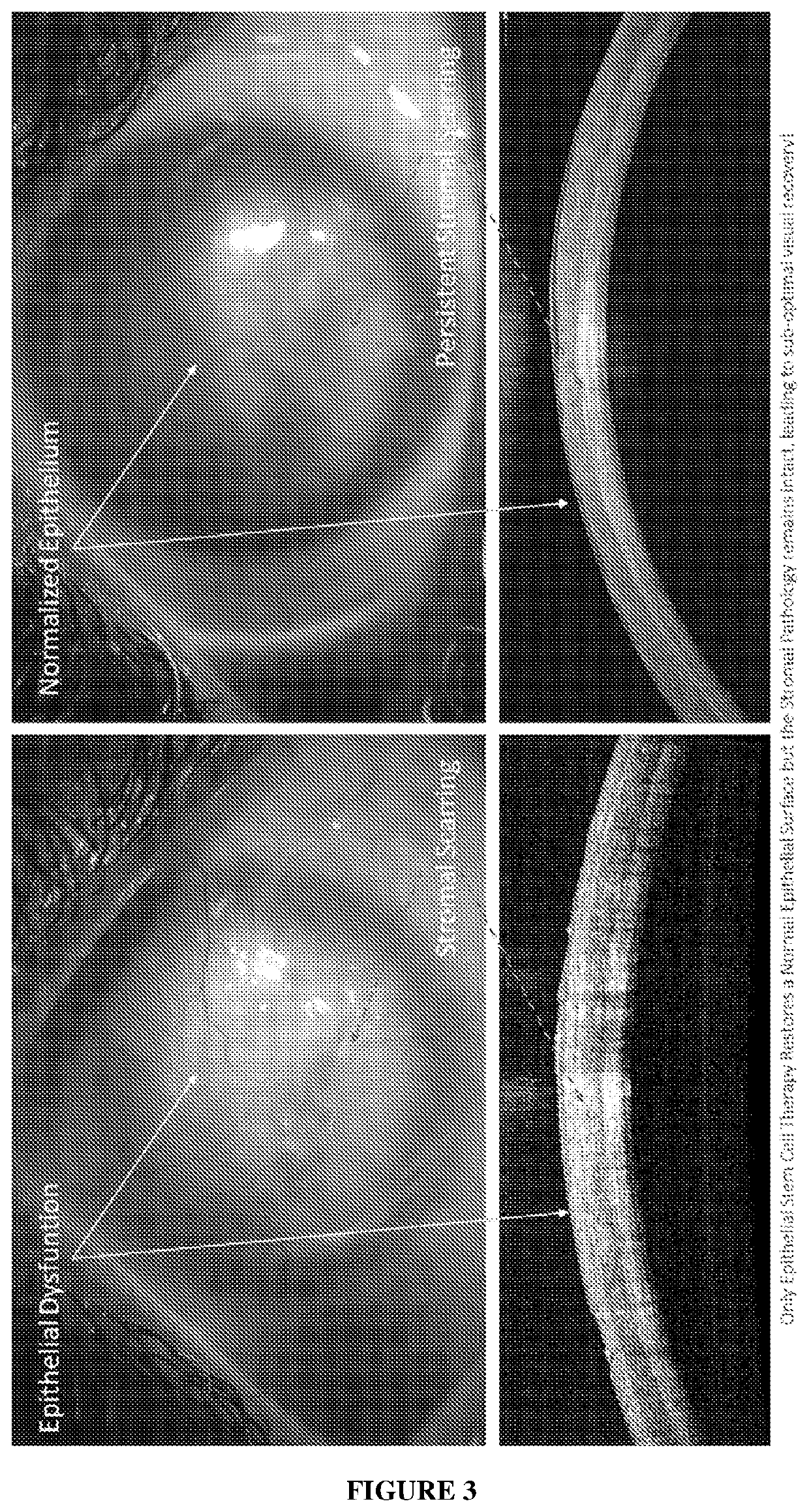
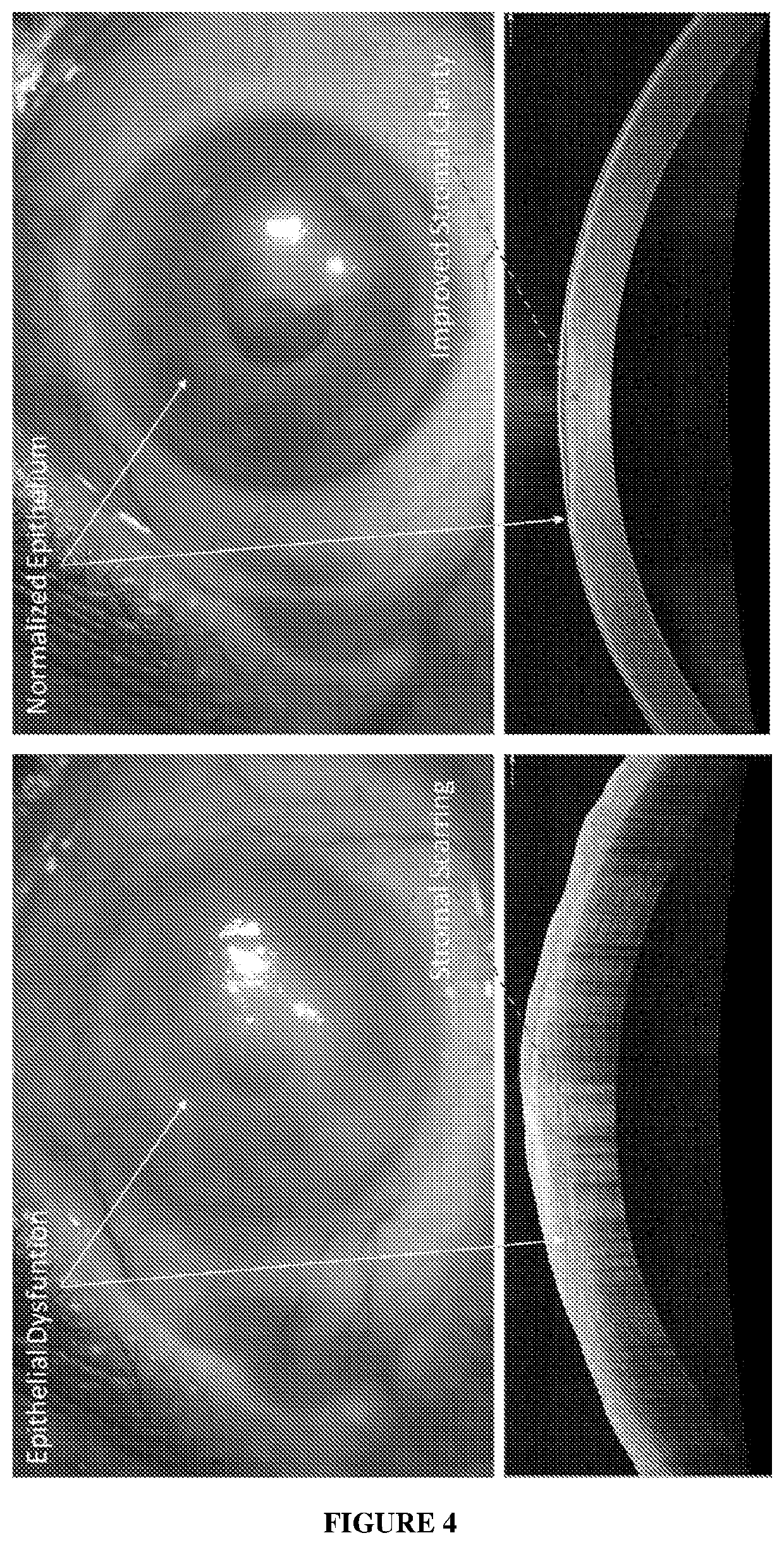
Cell Composition, Method of Production and its Use in Corneal Diseases
a cell composition and corneal disease technology, applied in the field of pharmaceutical compositions, can solve the problems of loss of corneal transparency, total blindness, deterioration of vision, etc., and achieve the effect of improving corneal clarity and visual recovery
- Summary
- Abstract
- Description
- Claims
- Application Information
AI Technical Summary
Benefits of technology
Problems solved by technology
Method used
Image


Examples
example 1
g Epithelial Stem Cells
[0090]Therapeutically accepted and serologically tested cadaveric corneas were obtained within four days of collection from the Ramayamma International Eye Bank (L. V. Prasad Eye Institute, Hyderabad, India). The corneas were washed with 1.25 mM penicillin-streptomycin (manufactured by Sigma-Aldrich®) followed by a wash with phosphate buffer saline (manufactured by Sigma-Aldrich®) at pH 7.4 for 3 minutes. It was followed by another wash with phosphate buffer saline.
[0091]Iris and endothelial layer were scrapped for visibility. Complete 360° limbal rims were isolated using a surgical blade in buffer saline and minced using a small, curved corneal scissors, in incomplete media (plain DMEM / F-12 media, manufactured by Lonza®).
[0092]The tiny limbal tissues pieces were subjected to collagenisation by adding 40μL of reconstituted Collagenase-IV to the incomplete media (Ser. No. 17 / 104,019, Thermofisher®) at the rate of 20 μL of Collagenase-IV per mL of incomplete med...
example 2
g Stromal Stem Cells
[0095]The epithelial stem cells (P0 culture) obtained in the previous example was further differentiated into stromal stem cells. Before the culturing, 1 mL of the spent media was collected into a sterile 1.5 mL microcentrifuge tube for mycoplasma contamination assay.
[0096]The media was discarded from the epithelial stem cell culture (P0 culture) and the cells were washed with phosphate buffer saline (manufactured by Sigma-Aldrich®) The cells were trypsinized by adding 1 mL TrypLE (manufactured by Thermofisher®) The flask was gently tapped 2-3 times and incubated at 37° C. for 2 minutes.
[0097]The trypsinized cells were transferred to a 15 mL centrifuge tube containing 1 mL complete media (plain DMEM / F-12 media, manufactured by Lonza®) The flask was washed with 2 mL phosphate buffer saline (manufactured by Sigma-Aldrich®) and added to the centrifuge tube. The cell culture was centrifuged at 1000 rpm for 3 minutes at 25° C. The supernatant was discarded, and the ce...
example 3
on of Cell Composition and Delivery of the Composition to the Patient
[0103]The epithelial stem cells and stromal stem cells as harvested from P0 and P3 cultures respectively were used for preparation of the multilayer cell composition and delivery to the patient.
[0104]The cell cultures were collected and checked for mycoplasma contamination assay.
[0105]Further, 10 μL of 4% Trypan blue stain was mixed with 10 μL of each of the cell suspension on a strip of parafilm to perform a cell count using Neubauer chamber to check the viability of the cell culture. The cell count in each of the cell culture must range between 4000 to 5000 cells per μL.
[0106]The supernatant of the stromal stem cells was discarded and 100 μL of stromal stem cells were taken. Similarly, supernatant of the epithelial stem cells was discarded and 200 μL of epithelial stem cells were added and mixed with the stromal cells to prepare the cell composition.
[0107]The ratio of the stromal stem cells and epithelial stem ce...
PUM
| Property | Measurement | Unit |
|---|---|---|
| Fraction | aaaaa | aaaaa |
| Fraction | aaaaa | aaaaa |
| Fraction | aaaaa | aaaaa |
Abstract
Description
Claims
Application Information
 - R&D
- Intellectual Property
- Life Sciences
- Materials
- Tech Scout
- Unparalleled Data Quality
- Higher Quality Content
- 60% Fewer Hallucinations
Browse by: Latest US Patents, China's latest patents, Technical Efficacy Thesaurus, Application Domain, Technology Topic, Popular Technical Reports.
© 2025 PatSnap. All rights reserved.Legal|Privacy policy|Modern Slavery Act Transparency Statement|Sitemap|About US| Contact US: help@patsnap.com