Method for isolating and detecting cancer stem cells

a cancer stem cell and isolating technology, applied in the field of isolating and detecting cancer stem cells, can solve the problem of faster implementation of existing methods

Pending Publication Date: 2021-02-25
CARCIDIAG BIOTECH
View PDF1 Cites 0 Cited by
  • Summary
  • Abstract
  • Description
  • Claims
  • Application Information

AI Technical Summary

Benefits of technology

[0018]The present invention relates to a specific method of detection since it recognizes only cancer stem cells of hormone-dependent cancer target organs and is therefore more efficient than conventional methods. In addition, its implementation is faster compared to existing methods, because the latter cannot be generalized due to their non-reproducibility and combine both stem and non-cancer stem cells. Hormone-dependent cancers are cancers sensitive to sex hormones. Hormone-dependent tumors mainly form in tissues whose functioning is normally regulated by hormones. The growth of tumors is stimulated by hormones, such as, for example, testosterone, a male sex hormone in the case of prostate cancer, or estrogen, a female sex hormone secreted by the ovaries, in the case of breast cancer.

Problems solved by technology

In addition, its implementation is faster compared to existing methods, because the latter cannot be generalized due to their non-reproducibility and combine both stem and non-cancer stem cells.

Method used


Image

  • Method for isolating and detecting cancer stem cells
  • Method for isolating and detecting cancer stem cells
  • Method for isolating and detecting cancer stem cells

Examples


example 1

for the Isolation of Cancer Stem Cells from Breast Cancer

[0295]I. Materials Required

[0296]Reagents and Materials[0297]Biotinylated individual lectin or mixture of biotinylated lectins specifically marking Cancer Stem Cells of hormone-dependent cancers, here breast cancer (prepared from individual lectins from Vector Laboratories and Emelca Biosciences)[0298]CELLection Biotin Binder kit (Invitrogen) containing magnetic beads coupled to streptavidin by a DNA bond[0299]Magnet

[0300]Buffers[0301]Versene (Invitrogen) comprising phosphate buffered saline (PBS) and EDTA[0302]Buffer 1: PBS (Phosphate buffered saline without Ca2+ and Mg2+) with 0.1% BSA (Bovine serum albumin), pH 7.4[0303]Buffer 2: PBS (Phosphate Buffer Saline without Ca2+ and Mg2+) with 0.1% BSA (Bovine Serum Albumin) and 0.6% sodium citrate[0304]Buffer 3: RPMI 1640 with 1% FCS (fetal calf serum), 1 mM CaCl2) and 5 mM MgCl2, pH 7.0-7.4.

[0305]II. Duration of the Experiment[0306]20 min to prepare cells[0307]20 min to label cel...

example 2

city Test

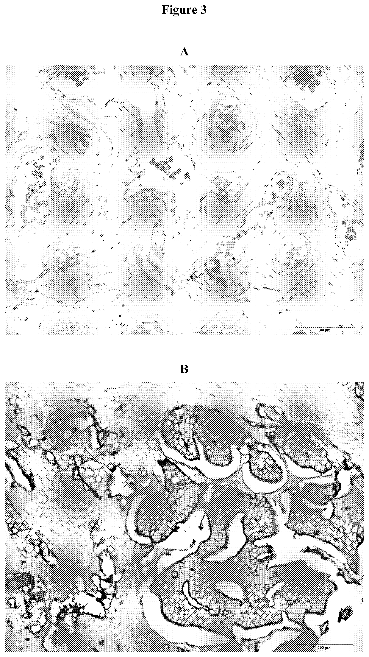
[0337]The objective of a clonogenicity test is to observe the capacity of cells to reform spheres (corresponding in the patient to the reform of a tumor mass) and therefore their proliferative capacity.

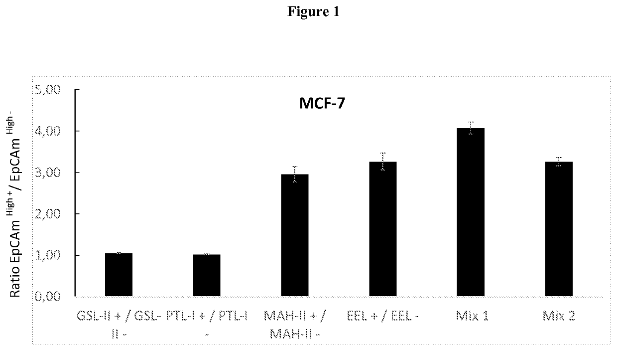
[0338]The clonogenicity test is in this example used to confirm the presence of cancer stem cells of hormone-dependent cancers and to quantify said cells in a sample after isolation of cancer stem cells of hormone-dependent cancers by the isolation method described herein. invention. It thus makes it possible to demonstrate the effectiveness of the isolation method according to the invention compared to a control sample not subjected to this method (unsorted cells).

[0339]The clonogenicity tests were carried out in a 6-well plate at a density of 500 cells / cm2 in a medium of RPMI composition (Gibco) supplemented with 50 units / ml of penicillin, 50 units / ml of streptomycin (Gibco) and 2.4 g / L of sodium bicarbonate, 1 M of HEPES buffer (Sigma Aldrich, Saint-Quentin-Fallavier, Fran...


PUM

PropertyMeasurementUnit
densityaaaaaaaaaa
weight ratioaaaaaaaaaa
thicknessaaaaaaaaaa

Abstract

Disclosed is the in vitro use of at least one lectin for marking cancer stem cells of hormone-dependent cancer target organs, selected from the lectins Maackia amurensis lectin II (MAH-II), Euonymus europaeus lectin (EEL), Psophocarpus tetragonolobus lectin I (PTL-I) and Griffonia simplicifolia lectin II (GSL-II), in particular at least two lectins selected from MAH-II, EEL, PTL-I and GSL-II, in particular the two lectins MAH-II and EEL, in order to obtain cancer stem cells of labeled hormone-dependent cancer target organs in a biological sample.

Description

CROSS-REFERENCE TO RELATED APPLICATIONS[0001]This application is the U.S. national phase of International Application No. PCT / FR2019 / 050516 filed Mar. 7, 2019 which designated the U.S. and claims priority to French Application No. 18 / 00202 filed Mar. 8, 2018, the entire contents of each of which are hereby incorporated by reference.BACKGROUND OF THE INVENTIONField of the Invention[0002]The present invention relates to a method of isolating and detecting cancer stem cells (CSCs) of hormone-dependent cancer target organs.[0003]The hormone-dependent cancer target organs are in particular the breast, uterus, prostate, ovaries, endometrium, thyroid and adrenal glands.Description of the Related Art[0004]Breast cancer is the leading cause of cancer death in France in women. It affects 48,000 people per year. Nearly 12,000 people per year die from this pathology with about one in 10 women affected during her lifetime. The 5-year survival rate is very low in the context of metastasized breas...

Claims


Application Information

Patent Timeline
Patent Type & Authority Applications(United States)
IPC IPC(8): A61K38/16G01N33/574C07K14/42C12N5/095A61K47/54A61K47/69
CPCA61K38/168G01N33/57484A61K47/6923C12N5/0695A61K47/557C07K14/42G01N33/5073G01N33/57407G01N33/57415G01N33/57469G01N33/57492G01N2333/42G01N2333/4724G01N2800/54
Inventor CARRE, VINCENTLACROIX, AURÉLIE
Owner CARCIDIAG BIOTECH
Why Eureka
Try Eureka