
Automated microscopic cell analysis

an automatic and cell technology, applied in the field of automatic microscopic cell analysis tasks, can solve the problems of time-consuming manual methods, laborious, limited accuracy, etc., and achieve the effects of saving time, improving sample preparation, and improving quantitative accuracy

Active Publication Date: 2021-02-11
MEDICA CORP
View PDF0 Cites 2 Cited by
  • Summary
  • Abstract
  • Description
  • Claims
  • Application Information

AI Technical Summary

Benefits of technology

The present invention is an apparatus for analyzing cells or particles in a biological sample. The technical effects of this invention include the ability to provide accurate counts of cells or particles without specialized knowledge or skills, in a fast and easy manner. Additionally, the invention allows for the analysis of small finger stick samples and pre-configured reagents and diluents. Furthermore, the invention creates a homogenous monolayer of cells and performs a Complete Blood Cell Analysis (CBC) in a few minutes. Additionally, the invention provides internal monitoring of the critical process steps of cell analysis to flag potentially erroneous results. The invention also enables the analysis of samples immediately after collection and avoids the need for transport or storage of samples in a laboratory.

Problems solved by technology

This manual method is time consuming, tedious, and requires a skilled technician to operate the microscope and to recognize the various types of cells, and is prone to error.
Its accuracy is limited by the number of cells counted and the uniformity of the monolayer formed by introduction of the diluted sample.
The analyzers are relatively large and expensive and require skilled operators for their use and maintenance.
This process adds costs and risk of erroneous results from transport, handling, labeling and transcription, as well as a time delay in obtaining the results.
Because Larsen's disclosure is directed to the analysis of a small sample of whole blood, errors in metering various volumes or in the mixing or sampling steps can significantly impact the accuracy of the results.
Maintaining uniformity and consistency of the 100 μm dimension of the channel height in a disposable cartridge is difficult to achieve in a low cost device.
In theory this approach has appeal, but the handling of undiluted whole blood is challenging.

Method used


Image

  • Automated microscopic cell analysis
  • Automated microscopic cell analysis
  • Automated microscopic cell analysis

Examples


example 1

on from Bright-Field and Fluorescent Optics

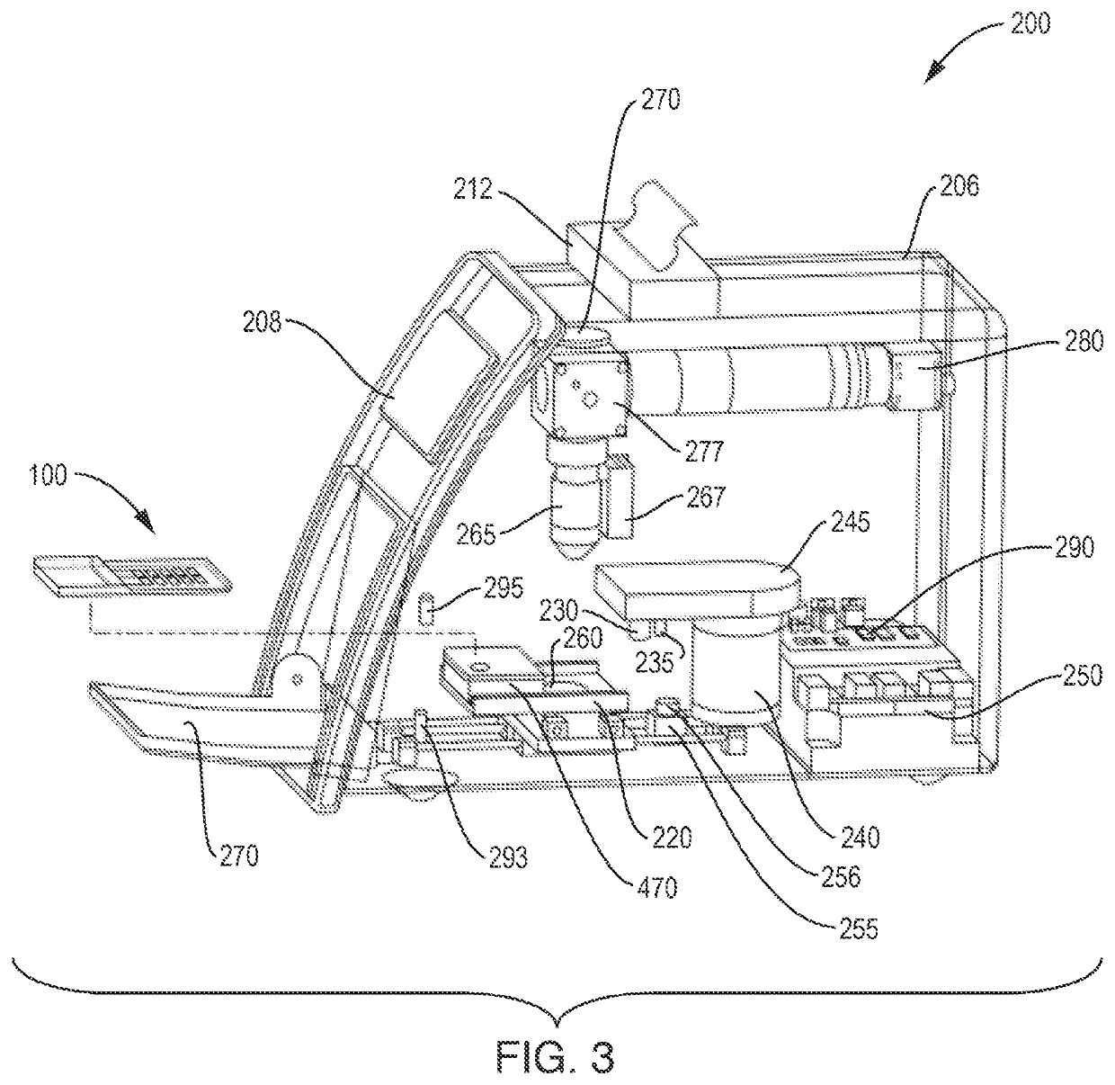
[0099]FIG. 16 shows images that were collected using test devices according to the present invention. A fluorescent stain Acridine Orange (AO) was used to differentially stain DNA and RNA of cells in a whole blood sample. The visual images of FIG. 16 were obtained using an Olympus 20×0.4 NA objective lens 265 and a Basler 5 MP digital camera 280. Excitation of the bright-field images in the second column was provided by white light bright-field source 260. Excitation of the fluorescent images in the third column was a 455 nm blue fluorescent light source 270.

[0100]White blood cells have significant RNA and DNA and therefore can be seen in the fluorescent images having green and orange structures. The size and shape of the green nuclear structure and overall size of the white cells can be used to differentiate them into sub-groups identified by name in the first column. Notably the basophil and eosinophil sub-groups of white cells have chara...

example 2

al Sampling of the Imaging Chamber

[0102]Table 1 illustrates a comparison of CBC parameters obtained according to the present invention and from an automated hematology analyzer.

TABLE 1Column1# pairsRBCsWBCsROIRBC / fRBC / f(%)-WBC / fWBC / f(%)RBC / WBC100% 9916245549251253818.7643.02100.01.342100.0479.1250%4958122966925351913.7642.5699.91.32598.7485.0825%24796230481285968.5643.28100.01.32798.9484.8610%992242197519373.5648.48100.81.390103.5466.66 5%496126186262197.2639.8299.51.32899.0481.63 1%100236836335.6664.61103.41.768131.7375.92 indicates data missing or illegible when filed

Sample: Low WBC count—approximately 2000 / uL (normal is 3,000-10,000 / uL).

Magnification: 20×

[0103]Number of images: approximately 10,000 bright-field and 10,000 fluorescent

Variable: Column 1—Percentage of total cells use in the calculation

Column # pairs—the number of pairs of images (bright-field plus fluorescent)

Column RBCs—total number of Red Blood Cells counted

Column WBCs—total number of White Blood Cells counted

Col...


PUM

PropertyMeasurementUnit
widthaaaaaaaaaa
widthaaaaaaaaaa
widthaaaaaaaaaa

Abstract

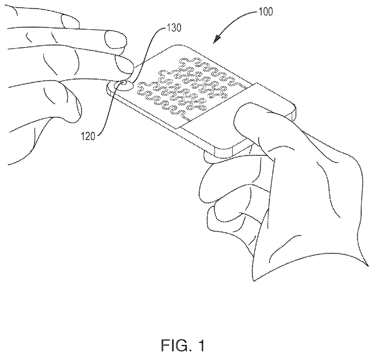
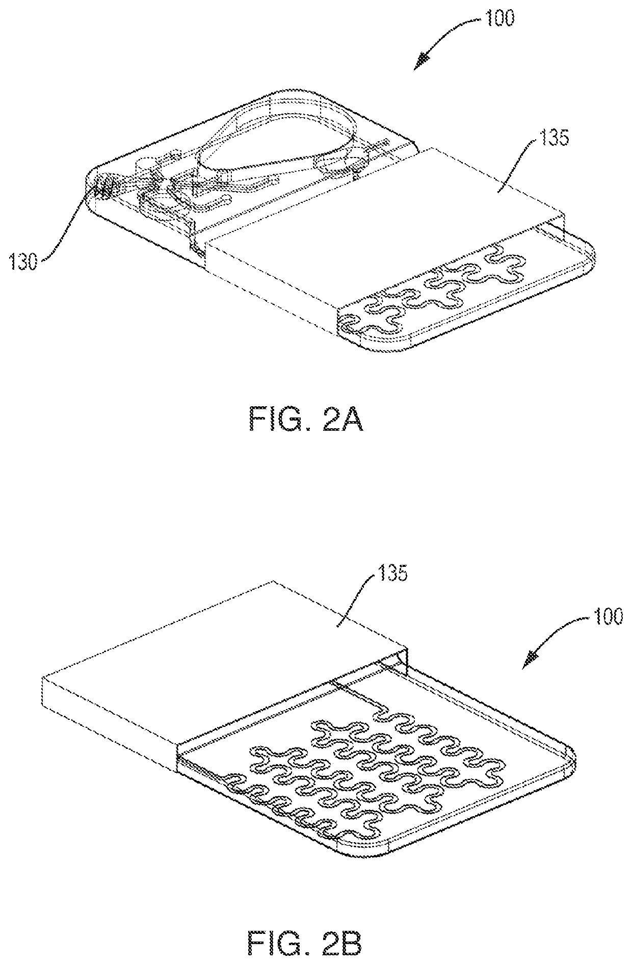
This disclosure describes single-use test cartridges, cell analyzer apparatus, and methods for automatically performing microscopic cell analysis tasks, such as counting and analyzing blood cells in biological samples. A small measured quantity of a biological sample, such as whole blood, is placed in a mixing bowl on the disposable test cartridge after being inserted into the cell analyzer. The analayzer also deposits a known amount of diluent / stain in the mixing bowl and mixes it with the blood. The analyzer takes a measured amount of the mixture and dispenses in a sample cup on the cartridge in fluid communication with an imaging chamber. The geometry of the imaging chamber is chosen to maintain the uniformity of the mixture, and to prevent cells from crowding or clumping as it is transferred into the imaging chamber by the analyzer. Images of all of the cellular components within the imaging chamber are counted and analyzed to obtain a complete blood count.

Description

CROSS-REFERENCE TO RELATED APPLICATIONS[0001]This application is a continuation of U.S. application Ser. No. 15 / 616,327, filed Jun. 7, 2017, which claims priority to U.S. provisional application No. 62 / 394,702, filed Sep. 14, 2016, and to U.S. provisional application No. 62 / 360,236, filed Jul. 8, 2016, and which is a continuation-in-part of U.S. application Ser. No. 15 / 221,285, filed Jul. 27, 2016, which is a continuation of U.S. application Ser. No. 15 / 017,498, filed Feb. 5, 2016, which is a continuation-in-part of U.S. application Ser. No. 14 / 947,971, filed Nov. 20, 2015, which claims priority to U.S. provisional application Ser. No. 62 / 138,359, filed Mar. 25, 2015, 62 / 113,360 filed Feb. 6, 2015, and 62 / 084,760, filed Nov. 26, 2014. All of the above-listed applications are herein incorporated by reference.FIELD OF THE INVENTION[0002]This invention pertains to analyzers and methods for automatically performing microscopic cell analysis tasks, such as counting blood cells in biologi...

Claims


Application Information

Patent Timeline
Patent Type & Authority Applications(United States)
IPC IPC(8): B01L3/00G01N15/14G06K9/00
CPCB01L3/502715G01N15/1434G01N2015/1006G06K9/00134G06K9/00147B01L3/502738G01N33/4915B01L3/502761B01L2200/04B01L2200/0605B01L2200/148B01L2300/0883B01L2400/0644G01N33/80G01N33/56972G01N1/30G01N2035/00158G01N15/1468G01N15/1456G01N15/1484G01N2015/1486G06V20/69G01N15/01G01N15/1433B01L2200/16B01L2400/0633B01L2200/0647B01L2200/027B01L2300/0627G01N33/5094G01N1/10G01N21/05G01N33/487G01N33/49G06V20/693G06V20/698
Inventor JONES, RONALDGROPPER, ADRIANHAGOPIAN, ROBERTROGERS, CHARLESVITELLA, THOMASCOTE, TYLERBARRY, DONALDOSTERLOH, DIRKYI, CHEN
Owner MEDICA CORP
Learn More