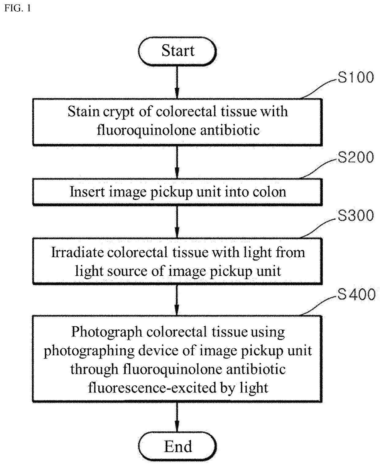
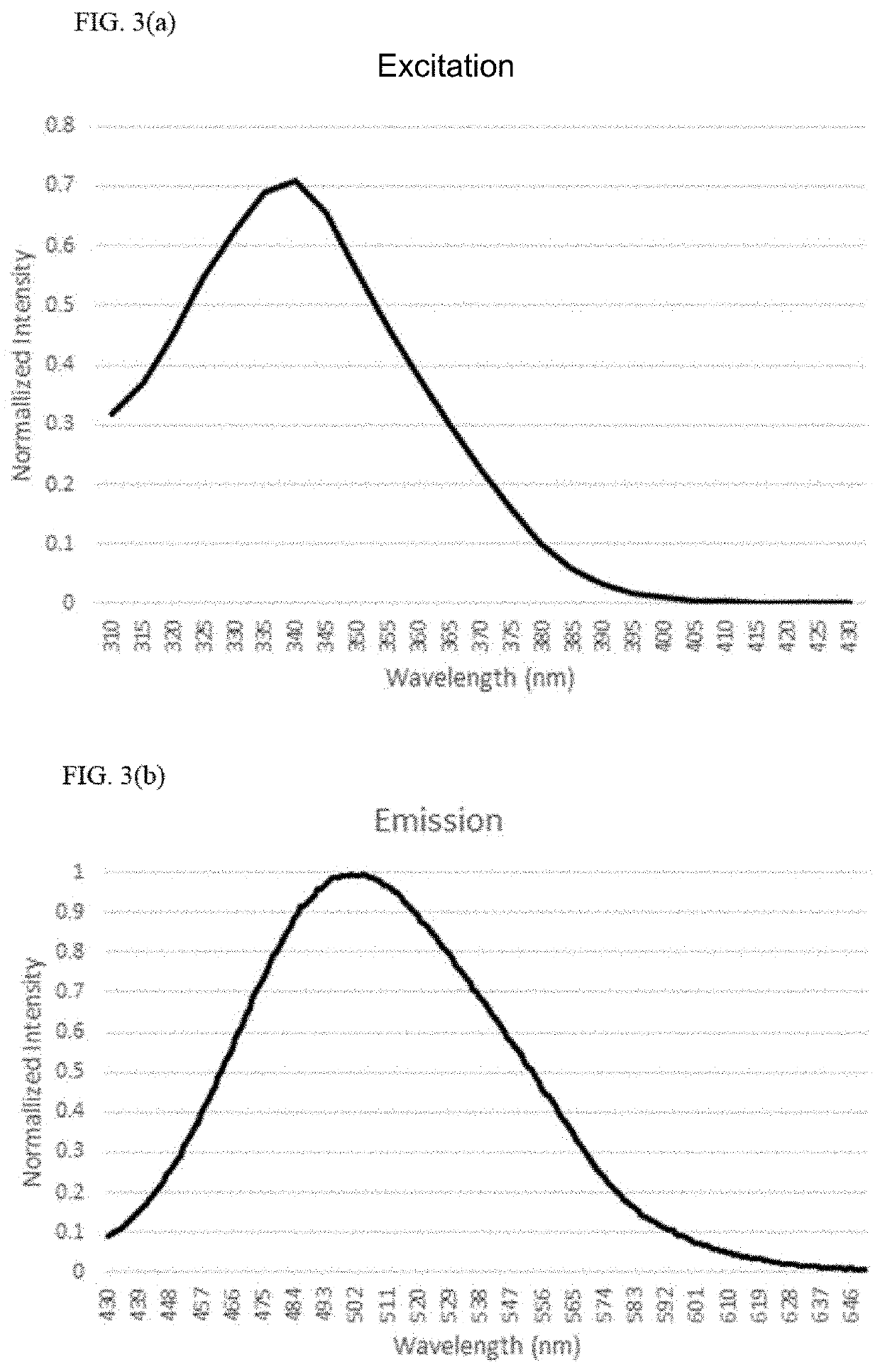
Fluorescence endoscopic method for visualization of colorectal tissue using fluoroquinolone antibiotics and method for diagnosis of lesions of colorectal tissue using the same
Active Publication Date: 2020-09-03
UNIV OF ULSAN FOUND FOR IND COOPERATION +3
View PDF0 Cites 0 Cited by
Summary
Abstract
Description
Claims
Application Information
AI Technical Summary
This helps you quickly interpret patents by identifying the three key elements:
Problems solved by technology
Method used
Benefits of technology
Problems solved by technology
First, since the colorectal tissue is stained with moxifloxacin, followed by irradiation of moxifloxacin with light in the visible region, high-speed direct photographing of the structure of the crypt is possible without damage to the colorectal tissue, thereby allowing quick and accurate diagnosis.
Second, since goblet cells of the colorectal tissue are stained with moxifloxacin to exhibit higher fluorescence intensity than surrounding cells, such that the crypt can be distinct, the crypt can be easily photographed even using a general fluorescence endoscope, which cannot provide 3D images.
Third, lesions of the colorectal tissue can be diagnosed based on numerical or morphological changes of goblet cells of the colorectal tissue.
Method used
Image
Click on the blue label to locate the original text in one second.
Reading with bidirectional positioning of images and text.
Examples
experimental example 1
Photographing of Extracted and Moxifloxacin-Stained Colorectal Tissue of Mouse
[0086]FIG. 4(a), FIG. 4(a1) and FIG. 4(b) are images of extracted and moxifloxacin-stained colorectal tissue of a mouse, obtained by the fluorescence endoscopic method for visualization of colorectal tissue using fluoroquinolone antibiotics according to the present invention.
[0087]FIG. 4(a) and FIG. 4(a1) are confocal fluorescence microscopy images of the moxifloxacin-stained colorectal tissue of the mouse. Referring to FIG. 4(a) and FIG. 4(a1), goblet cells of the colorectal tissue stained with moxifloxacin were fluorescence-excited and emitted light, whereby the crypt and goblet cells of the colorectal tissue could be observed.
[0088]FIG. 4(b) is a high-magnification fluorescence microscopy image of the moxifloxacin-stained colorectal tissue of the mouse. Referring to FIG. 4(b), goblet cells of the colorectal tissue stained with moxifloxacin were fluorescence-excited and emitted light, whereby the crypt a...
experimental example 2
Photographing of Moxifloxacin-Stained Colorectal Tissue of Living Mouse
[0090]FIG. 5(a), FIG. 5(a1), FIG. 5(b), and FIG. 5(b1) are images of moxifloxacin-stained colorectal tissue of a living mouse, obtained by the fluorescence endoscopic method for visualization of colorectal tissue using fluoroquinolone antibiotics according to the present invention.
[0091]FIG. 5(a) is a confocal fluorescence microscopy image of the moxifloxacin-stained colorectal tissue of the mouse and FIG. 5(a1) is an enlarged (high-magnification) image of a part of FIG. 5(a). Referring to FIG. 5(a) and FIG. 5(a1), goblet cells of the colorectal tissue stained with moxifloxacin was fluorescence-excited and emitted light, whereby the crypt and goblet cells of the colorectal tissue could be observed.
[0092]FIG. 5(b) is a high-magnification fluorescence microscopy image of the moxifloxacin-stained colorectal tissue of the mouse and FIG. 5(b1) is an enlarged (high-magnification) image of a part of FIG. 5(b). Referring...
experimental example 3
Photographing of Moxifloxacin-Stained Colorectal Tissue of Living Mouse Ingesting DSS
[0094]FIG. 6(a), FIG. 6(a1), FIG. 6(b), and FIG. 6(b1) are images of moxifloxacin-stained colorectal tissue of a mouse ingesting dextran sodium sulfate (DSS), obtained by the fluorescence endoscopic method for visualization of colorectal tissue using fluoroquinolone antibiotics according to the present invention.
[0095]FIG. 6(a) is a confocal fluorescence microscopy image of the moxifloxacin-stained colorectal tissue of the mouse and FIG. 6(a1) is an enlarged (high-magnification) image of a part of FIG. 6(a). Referring to FIG. 6(a) and FIG. 6(a1), it could be observed that, in the colorectal tissue of the mouse ingesting DSS, there was no light emission from cells due to collapse of the crypt or destruction of the goblet cells caused by DSS.
[0096]FIG. 6(b) is a high-magnification fluorescence microscopy image of the moxifloxacin-stained colorectal tissue of the mouse and FIG. 6(b1) is an enlarged (hi...
PUM
Property
Measurement
Unit
wavelength band
aaaaa
aaaaa
wavelength
aaaaa
aaaaa
fluorescence
aaaaa
aaaaa
Abstract
A fluorescence endoscopic method for visualization of colorectal tissue using a fluoroquinolone antibiotic and a method for diagnosis of lesions of colorectal tissue using the same. The fluorescence endoscopic method for visualization of colorectal tissue includes staining goblet cells of the colorectal tissue with moxifloxacin, which is a fluoroquinolone antibiotic, and exciting the stained goblet cells with single photons in the near UV region or in the visible region, followed by fluorescence endoscopic photographing of the goblet cells, thereby enabling acquisition of morphological information on living tissue without damage to or destruction of the colorectal tissue, while allowing diagnosis of lesions of the colon based on the morphological information on living tissue such as information on the distribution of the goblet cells. Specifically, the fluorescence endoscopic method includes: a colorectal tissue staining step in which the crypt of the colorectal tissue is stained with a fluoroquinolone antibiotic; an image pickup unit insertion step in which an image pickup unit is inserted into the colon; a light irradiation step in which the colorectal tissue is irradiated with excitation light from a light source of the image pickup unit; a colorectal tissue photographing step in which the colorectal tissue is photographed using a photographing device of the image pickup unit through the fluoroquinolone antibiotic fluorescence-excited by light in the light irradiation step, wherein, in the colorectal tissue staining step, goblet cells of the crypt are stained with the fluoroquinolone antibiotic; in the light irradiation step, the light source emits single photons as excitation light inducing fluorescence of the fluoroquinolone antibiotic, the excitation light from the light source having a wavelength band within the near UV and visible regions; and, in the image pickup unit insertion step, the image pickup unit is a general fluorescence endoscope or a confocal endoscopic microscope.
Description
STATEMENT REGARDING RESEARCH[0001]The research related to this disclosure was supported by the projects listed below:[0002][Project number] 1415157444[0003][Ministry] Ministry of Trade, Industry and Energy[0004][Management agency] Korea Evaluation Institute of Industrial Technology[0005][Program name] Industrial Technology Innovation Program (10063408) by the Ministry of Trade, Industry & Energe (MOTIE, Korea)[0006][Project name] Development of channel-inserted convergence smart endoscopic system based on multispectral fluorescence imaging for precise diagnosis of digestive diseases[0007][Contribution ratio] 20%[0008][Supervision institution] Asan Medical Center[0009][Period] Jan. 1, 2018˜Dec. 31, 2018[0010][Project number] 1711068372[0011][Ministry] Ministry of Science and ICT[0012][Management Agency] National Research Foundation of Korea[0013][Program name] Korea-Sweden Research Cooperation Program (2017R1A2A1A18070960) of the National Research Foundation (NRF) funded by the Korea...
Claims
Application Information
Patent Timeline
Application Date:The date an application was filed.
Publication Date:The date a patent or application was officially published.
First Publication Date:The earliest publication date of a patent with the same application number.
Issue Date:Publication date of the patent grant document.
PCT Entry Date:The Entry date of PCT National Phase.
Estimated Expiry Date:The statutory expiry date of a patent right according to the Patent Law, and it is the longest term of protection that the patent right can achieve without the termination of the patent right due to other reasons(Term extension factor has been taken into account ).
Invalid Date:Actual expiry date is based on effective date or publication date of legal transaction data of invalid patent.
Patent Type & Authority Applications(United States)