Devices, systems and methods for biomarker analysis
- Summary
- Abstract
- Description
- Claims
- Application Information
AI Technical Summary
Benefits of technology
Problems solved by technology
Method used
Image
Examples
example 1
r Analysis of Cell-Free Nucleic Acids from Whole Blood
[0315]A device for purifying separating plasma from maternal whole blood for the purpose of analyzing cell-free fetal nucleic acids was constructed. The device consists of 6 layers. From bottom to top these are:
[0316](1) Lower Adhesive Sheet
[0317](2) Lower Separation Disc: 16 mm diameter disc of adhesive sheet material (polymer material that is inert to DNA or Plasma) with glue on the side facing the Lower Adhesive Sheet
[0318](3) Polyethersulfone (PES) membrane, various sizes, typically between 6 and 16 mm, preferred design features 10 mm PES membrane. The membrane serves as wicking material which attracts the plasma from the filter through capillary force.
[0319](4) Filter Disc (e.g., Pall Vivid™ Membrane), 16 mm diameter, rough side facing up, shiny side facing the PES membrane.
[0320](5) Upper Separation Disc: same material as Lower Separation Disc, size 12 or 14 mm diameter, containing a 4 mm hole in the center. When using adhe...
example 2
r Analysis of Fetal Cell-Free Nucleic Acids from Maternal Blood
[0325]The device consists of multiple layers as exemplified in Example 1.
[0326]Application of blood and filtration to the device occurs as follows:
[0327]40 μl to 60 μl of whole blood is applied to the center of the device through the hole in the Upper Adhesive Sheet and the hole in the Upper Separation Disc. The blood distributes centripetally throughout the Filter Disc by capillary forces. Plasma is also wicked through the Filter Disc into the PES membrane by capillary forces. After about two minutes, the maximum amount of plasma has been transferred into the PES membrane.
[0328]The PES membrane containing cell-free nucleic acids is recovered as follows:
[0329]The device is cut out around the edges of the PES membrane. The membrane separates easily from the Filter and the Lower Disc.
[0330]DNA is eluted from the membrane as follows:
[0331]The PES membrane containing the plasma is transferred into an Eppendorf tube (0.5 ml) ...
example 3
of Human Y Chromosome DNA Using Recombinase Polymerase Amplification
[0333]Amplification and detection of human Y chromosome DNA in plasma samples were carried out by the following various methods:
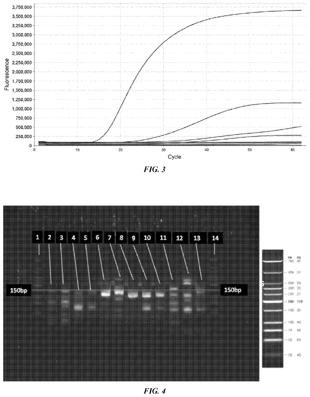
[0334]Detection of Y Chromosome targets using RPA and polyacrylamide gel electrophoresis (PAGE):
[0335]Recombinase polymerase amplification of 50 ng (15151 copies) of male genomic DNA was conducted using the TwistAmp Basic Kit (TwistDx, Cambridge, UK) following the standard protocol. Briefly, 29.5 μl of Rehydration Buffer was combined with 3 μl of each amplification primer (10 μM) (IDT, Coralville, Iowa), 2 μl of water and 10 μl of DNA template. 47.5 μl of this mixture was mixed with the lyophilized RPA enzymes as provided (uvsX, uvsY, gp32, Bsu). Following resuspension of the lyophilized RPA enzymes the reaction mixture was added to 2.5 μl of 280 mM magnesium acetate and mixed thoroughly to activate the RPA reaction. The reaction was incubated at 37 degrees Celsius for 20 minutes with agita...
PUM
| Property | Measurement | Unit |
|---|---|---|
| Fraction | aaaaa | aaaaa |
| Fraction | aaaaa | aaaaa |
| Pore size | aaaaa | aaaaa |
Abstract
Description
Claims
Application Information
 - R&D
- Intellectual Property
- Life Sciences
- Materials
- Tech Scout
- Unparalleled Data Quality
- Higher Quality Content
- 60% Fewer Hallucinations
Browse by: Latest US Patents, China's latest patents, Technical Efficacy Thesaurus, Application Domain, Technology Topic, Popular Technical Reports.
© 2025 PatSnap. All rights reserved.Legal|Privacy policy|Modern Slavery Act Transparency Statement|Sitemap|About US| Contact US: help@patsnap.com


