
Histopathological image analysis

a histopathologist and image technology, applied in the field of histopathological image analysis, can solve the problems of difficult or impossible for a histopathologist to many features of interest are still difficult or impossible for a histopathologist to see or accurately quantify with h&e staining, and the ihc technique may be expensive, time-consuming, and/or difficult to implement. achieve the effect of convenient, quick and easy us

Active Publication Date: 2020-07-16
ROOM4 GROUP
View PDF0 Cites 15 Cited by
  • Summary
  • Abstract
  • Description
  • Claims
  • Application Information

AI Technical Summary

Benefits of technology

This patent describes a method for analyzing histological specimens using a machine-learning algorithm. The algorithm can predict the appearance of a second image of a specimen if it had been stained with a different marker. The method can also use a faster, easier, or cheaper marker to obtain simulated results that are comparable to using the second marker. The second specimens can be the same or adjacent sections of biological tissue. Dividing the training images into tiles increases the amount of training data and controls the size and complexity of the algorithm. The algorithm can be trained to classify each pixel of a tile as exhibiting the histological feature of interest, or not. The method can use H&E dye, which is widely used in the art to stain histological specimens.

Problems solved by technology

Unfortunately, many features of interest are still difficult or impossible for a histopathologist to see or accurately quantify with H&E staining.
However, IHC is not without drawbacks.
IHC techniques may be expensive, time-consuming, and / or difficult to implement.

Method used


Image

  • Histopathological image analysis
  • Histopathological image analysis
  • Histopathological image analysis

Examples


Embodiment Construction

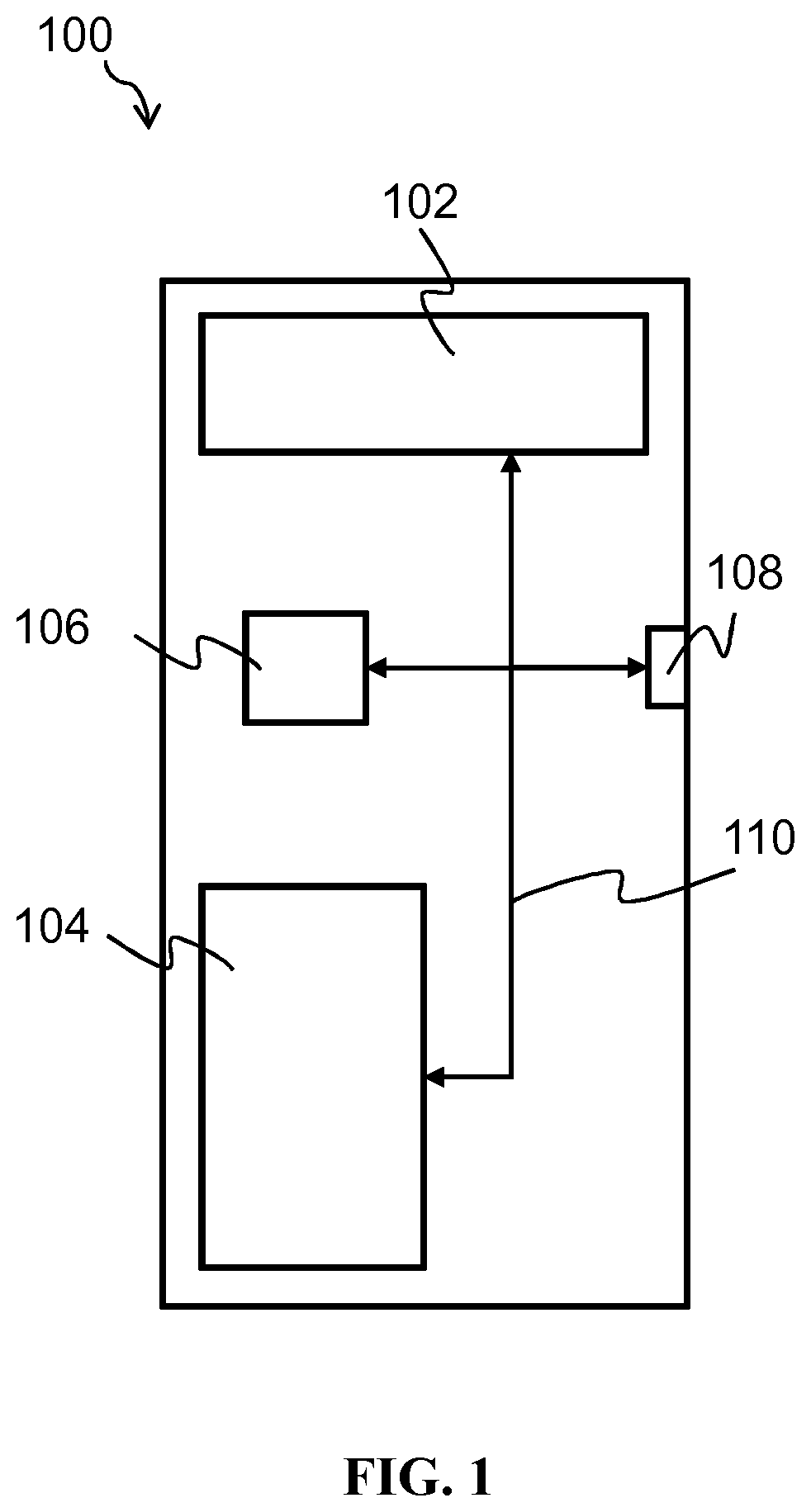
[0082]FIG. 1 of the accompanying drawings schematically illustrates an exemplary computer system 100 upon which embodiments of the present invention may run. The exemplary computer system 100 comprises a computer-readable storage medium 102, a memory 104, a processor 106 and one or more interfaces 108, which are all linked together over one or more communication busses 110. The exemplary computer system 100 may take the form of a conventional computer system, such as, for example, a desktop computer, a personal computer, a laptop, a tablet, a smart phone, a smart watch, a virtual reality headset, a server, a mainframe computer, and so on. In some embodiments, it may be embedded in a microscopy apparatus, such as a virtual slide microscope capable of whole slide imaging.

[0083]The computer-readable storage medium 102 and / or the memory 104 may store one or more computer programs (or software or code) and / or data. The computer programs stored in the computer-readable storage medium 102 ...


PUM

PropertyMeasurementUnit
fluorescenceaaaaaaaaaa
microscopic structureaaaaaaaaaa
transparentaaaaaaaaaa

Abstract

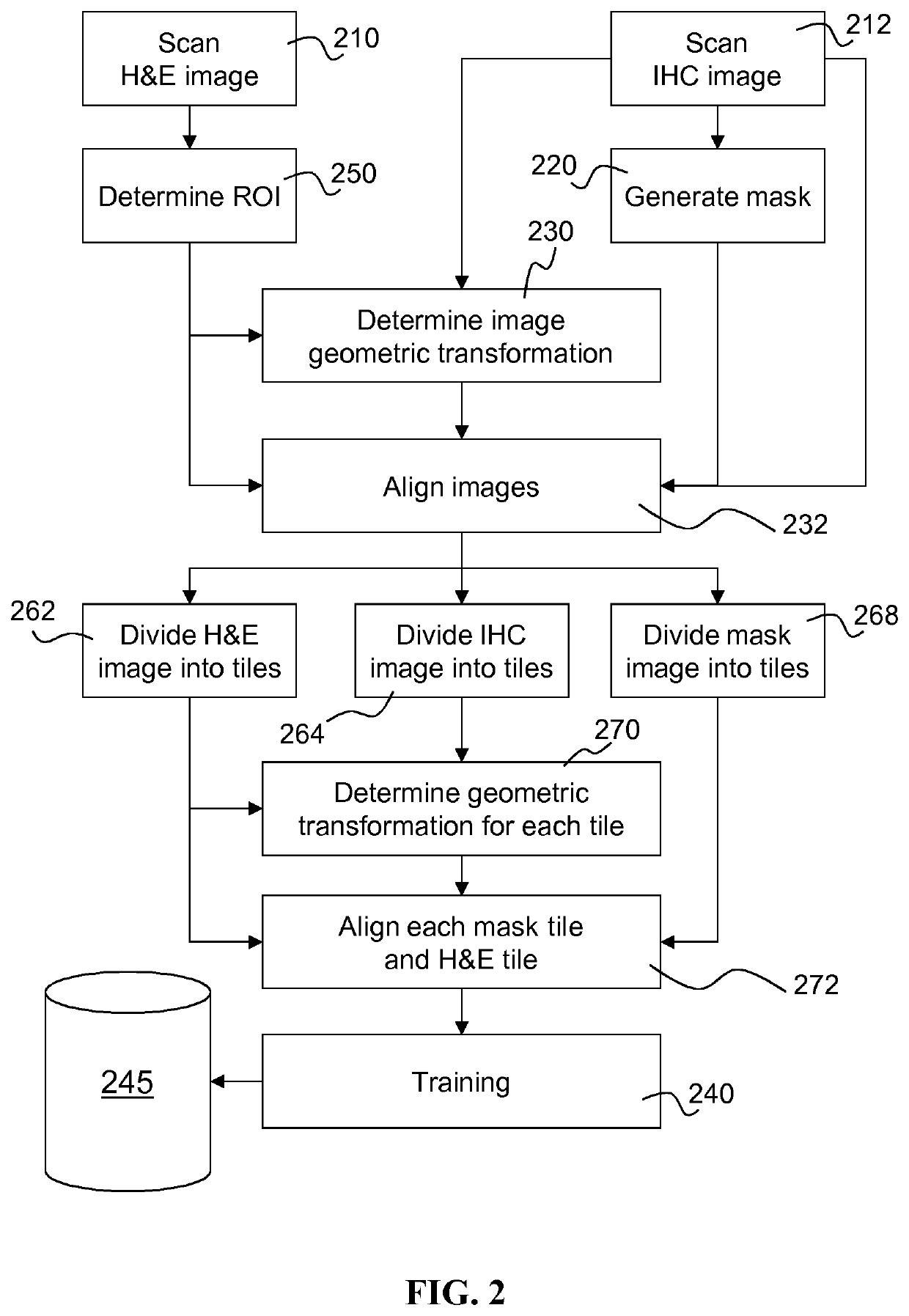
An apparatus and computer-implemented method for training a machine-learning algorithm to perform histopathological analysis is disclosed. The method comprises obtaining (210) a plurality of first microscopic images of first histological specimens that have been stained with a first marker; and obtaining (212), a respective plurality of second microscopic images of second histological specimens that have been stained with a second, different marker. The method further comprises obtaining (220) a respective plurality of mask images generated for the second microscopic images, each mask image identifying a histological feature of interest highlighted in the respective second microscopic image by the second marker. The method comprises training (240) the machine-learning algorithm to predict, from a first microscopic image, a histological feature of interest that would be highlighted in the same specimen by the second marker. Also disclosed is an apparatus and computer-implemented method for histopathological analysis using the trained machine-learning algorithm.

Description

FIELD OF THE INVENTION[0001]This invention relates to analysis of histological images. It relates in particular to using a machine-learning algorithm to perform such analysis and also to training the machine-learning algorithm to perform the analysis.BACKGROUND OF THE INVENTION[0002]As used herein, a “histological image” refers to an image showing the microscopic structure of organic tissue. A “histological feature of interest” means a feature of this microscopic structure. The feature may be of interest for diagnostic or therapeutic purposes, or for scientific research, for instance. One example of a histological feature of interest is stroma—the connective and supportive framework of a tissue. Identification of stroma in biopsy specimens may be useful for cancer diagnosis and determining prognosis. Another example of a histological feature of interest is chromatin. In general, methods allowing the characterisation of cell nuclei have drug discovery, clinical and other medical appl...

Claims


Application Information

Patent Timeline
Patent Type & Authority Applications(United States)
IPC IPC(8): G06N3/08G06N20/00G06T7/00G06K9/00G06K9/46G06K9/32G01N33/53
CPCG06K9/0014G06T2207/20084G06N3/08G06N20/00G06T2207/20021G01N33/53G06T2207/10056G06T7/0014G06K9/4671G06K9/32G06K9/00134G06T2207/20081G06T7/0012G06T2207/10064G06T2207/30024G06V20/695G06T7/337A61B5/0033A61B6/48A61B6/50G06T2207/30004G06V20/693
Inventor MADDISON, JOHN ROBERTDANIELSEN, HAVARD
Owner ROOM4 GROUP
Learn More