
Medical diagnosis support device, endoscope system, and medical diagnosis support method

a medical diagnosis and support device technology, applied in the recognition of medical/anatomical patterns, instruments, image enhancement, etc., can solve the problems of inconvenient use, observation or diagnosis may be hindered, etc., and achieve the effect of eliminating anxiety and facilitating the setting of waiting tim

Active Publication Date: 2020-05-07
FUJIFILM CORP
View PDF0 Cites 4 Cited by
  • Summary
  • Abstract
  • Description
  • Claims
  • Application Information

AI Technical Summary

Benefits of technology

The invention is a medical diagnosis support device and an endoscope system that can detect and discriminate medical images. The device has a waiting time before displaying the results of detection or discrimination based on the user's settings. This prevents the user from feeling inconvenien or hindering observation or diagnosis. The device can also display information representing that the result is not displayed, which helps to eliminate user anxiety. The endoscope system includes the medical diagnosis support device and an endoscope that can be inserted into an object to be examined, and a light source device that can switch between different illumination lights based on the detection and discrimination results. This improves the visibility of the recognition result of the medical image.

Problems solved by technology

For this reason, there is a concern that a user may feel inconvenient and observation or diagnosis may be hindered.

Method used


Image

  • Medical diagnosis support device, endoscope system, and medical diagnosis support method
  • Medical diagnosis support device, endoscope system, and medical diagnosis support method
  • Medical diagnosis support device, endoscope system, and medical diagnosis support method

Examples


first embodiment

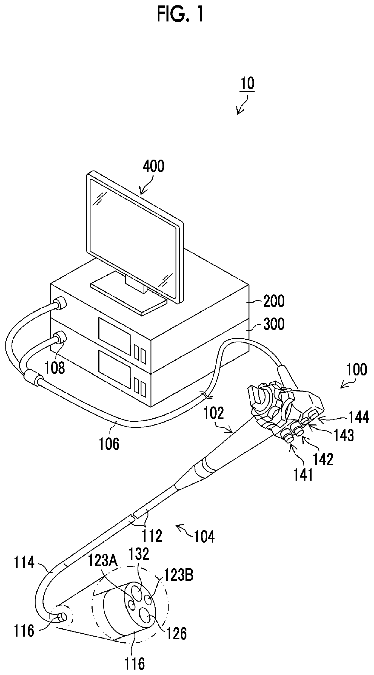
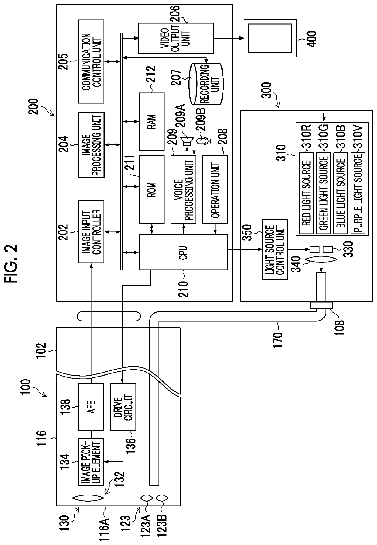
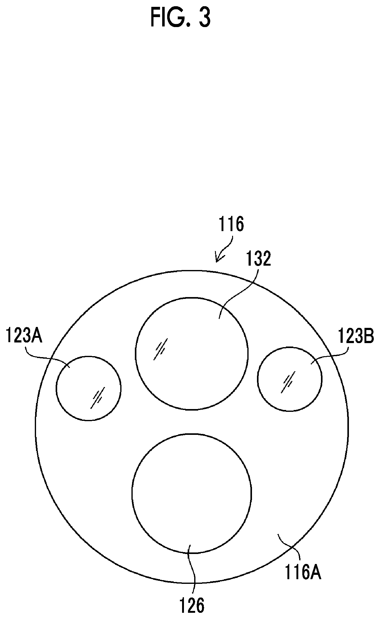
[0048]FIG. 1 is a diagram showing the appearance of an endoscope system 10 (a medical-use image processing device, a medical image processing device, a medical diagnosis support device, an endoscope system) according to a first embodiment, and FIG. 2 is a block diagram showing the main configuration of the endoscope system 10. As shown in FIGS. 1 and 2, the endoscope system 10 includes an endoscope 100 (an endoscope, an endoscope body), a processor 200 (a processor, an image processing device, a medical image processing device, a medical diagnosis support device), a light source device 300 (light source device), and a monitor 400 (display device).

[0049]Configuration of Endoscope

[0050]The endoscope 100 comprises a hand operation part 102 (hand operation part) and an insertion part 104 (an insertion part) connected to the hand operation part 102. An operator (user) grips and operates the hand operation part 102, inserts the insertion part 104 into an object to be examined (living body...


PUM


Abstract

An object of the invention is to provide a medical diagnosis support device, an endoscope system, and a medical diagnosis support method where the visibility of a recognition result of a medical image is high.A medical diagnosis support device according to an aspect of the invention includes an image acquisition section that acquires medical images in time series, a detection section that detects a region of interest included in the medical images, a discrimination section that performs the discrimination of the medical images, a display control section that causes a display device to display any one of a result of the detection or a result of the discrimination, and a setting section that sets a waiting time required until the display is performed after the detection or the discrimination is performed. In a case where an object to be displayed is switched between the result of the detection and the result of the discrimination, the display control section causes the result of the detection or the discrimination to be displayed when the waiting time has passed after the detection or the discrimination is performed.

Description

CROSS-REFERENCE TO RELATED APPLICATIONS[0001]The present application claims priority under 35 U.S.C. § 119 to Japanese Patent Application No. 2018-207122, filed on Nov. 2, 2018. The above application is hereby expressly incorporated by reference, in its entirety, into the present application.BACKGROUND OF THE INVENTION1. Field of the Invention[0002]The present invention relates to a medical diagnosis support device, an endoscope system, and a medical diagnosis support method that perform the recognition of a medical image and display the result thereof.2. Description of the Related Art[0003]In diagnosis support using a medical image, there is a case where the recognition of an image is performed and the result thereof is displayed on a display device. For example, a recognition result is displayed through the emphasis display of a region of interest detected from an image, but a technique for delaying a timing where such a display is performed is known. For example, WO17 / 203560A dis...

Claims


Application Information

Patent Timeline
IPC IPC(8): A61B1/00G06K9/32A61B1/06G06V10/25G06V10/764
CPCG06K9/3233G06K2209/05A61B1/00006A61B1/0638A61B1/0005A61B1/043G06T7/0012G06T2207/20084G06V10/25G06V2201/03G06V10/82A61B1/000094A61B1/000096G06V10/764
Inventor ENDO, MAIKO
Owner FUJIFILM CORP
Learn More