Method for assessing corneal tissue quality and endothelial cell density and morphology

a corneal tissue and endothelial cell technology, applied in the field of corneal tissue quality assessment, can solve the problems of difficult analysis, inability to achieve satisfactory specular reflection or cell border definition of the percentage of human donor cornea endothelia, and achieve the effect of improving tissue quality assessment, reducing analysis time, and increasing endothelial cell border definition

Inactive Publication Date: 2019-10-03
CORNEAGEN INC
View PDF0 Cites 0 Cited by
  • Summary
  • Abstract
  • Description
  • Claims
  • Application Information

AI Technical Summary

Benefits of technology

[0011]First, the methods provide for a decreased timeframe for analysis of human donor corneas after cold storage. The traditional process of free convection at room temperature takes 2-3 hours on average before the donor cornea's endothelium is analyzable. Surprisingly, using Applicants' method of convection in / at an environmental temperature of up to about 37° C., that timeframe is reduced to, for example, about 0.75-1.5 hours on average, as disclosed and supported herein.
[0012]Second, the methods provide for increased endothelial cell border definition. As stated above, it is generally recognized in the art that even warming of corneas above room temperature in hypothermic storage medium has a deleterious effect on cell morphology and that folds induced by swelling of corneal tissue at such elevated temperatures results in endothelial cell damage and some cell loss. Human donor cornea endothelia are thus typically analyzed at room temperature, but where it is commonly difficult to analyze due to poorly defined cell borders. Applicants have surprising found that incubating cornea at temperatures above room temperature, and raising the temperature of the corneal tissue above room temperature, results in substantially improved tissue quality assessment. Without being bound by mechanism, at temperatures above room temperature and up to about natural body temperature (e.g., about 37° C.) the donor cornea endothelia appear to respond more typical of in vivo cells, creating better resolution of the cell borders. Moreover, with better defined borders and better specular reflection, a larger quantity of cells are elucidated, resulting in a higher and more accurate cell density analysis, as disclosed and supported herein in the working examples. Applicants, therefore, despite the conventional wisdom that incubation of corneal tissue after hypothermic storage should not exceed room temperature (to avoid tissue damage), have found that incubating corneal tissue in hypothermic corneal storage media to and / or at a temperature above ambient temperature (e.g., to and / or at a temperature approximating body temperature or approximating the cornea's natural functional temperature), results in substantially improved tissue quality assessment, while maintaining tissue quality and viability for transplantation. The discovery of the capacity of the tissue to maintain such quality and viability at tissue analysis above ambient temperature, and even after one or more periods of cold storage (e.g., at a temperature in the range of 2-8° C.), provides for new and useful methods over the prior art, as claimed herein.

Problems solved by technology

As stated above, it is generally recognized in the art that even warming of corneas above room temperature in hypothermic storage medium has a deleterious effect on cell morphology and that folds induced by swelling of corneal tissue at such elevated temperatures results in endothelial cell damage and some cell loss.
Human donor cornea endothelia are thus typically analyzed at room temperature, but where it is commonly difficult to analyze due to poorly defined cell borders.
According to particular aspects, a percentage of human donor cornea endothelia are not able to achieve satisfactory specular reflection or definition of cell borders at room temperature.

Method used


Image

  • Method for assessing corneal tissue quality and endothelial cell density and morphology
  • Method for assessing corneal tissue quality and endothelial cell density and morphology
  • Method for assessing corneal tissue quality and endothelial cell density and morphology

Examples


example 1

Exemplary Materials

[0051]Corneal Tissue.

[0052]Human donor corneal-scleral rims were used.

[0053]Hypothermic Storage Media.

[0054]Optisol™ GS was used for the present working examples experiments. Other exemplary media that Applicant's incubation technique encompasses are, e.g., Life4C.™, McCarey-Kaufman™ Media, EUSOL-C™, Dexsol™, K-Sol®, CSM™, Chen Medium™, Cornisol™, Steinhardt Media™, Likorol™, etc.

[0055]Corneal Preservation Chamber.

[0056]Exemplary corneal preservation chambers include, but are not limited to, for example, Krolman Viewing Chamber (as used in the present working examples), Bausch&Lomb Viewing Chamber, Numedis Transend Chamber, Alcon Viewing Chamber. Preservation in the original media chamber is also an option.

[0057]Specular Microscope.

[0058]HAI CAS EB-3000xyz (used for our experiments), Konan EB-10, or other suitable specular microscope.

[0059]Incubator.

[0060]Thermo-Scientific Heratherm Compact Incubator, or other suitable incubator

[0061]Slit-Lamp Microscope.

[0062]A T...

example 2

Exemplary Re-Evaluation, and Initial Evaluation Methods were Successfully Applied

[0063]EyeBank Specular Microscopy.

[0064]An overview of EyeBank Specular Microscopy is available on-line at “slideshare.net / EBAICME / eye-bank-specular-microscopy”.

[0065]Both re-evaluation and initial evaluation were performed as follows:

[0066]A—Re-Evaluation by Incubation at 34° C. in Optisol™ GS after an Initial Evaluation at Room Temperature:

[0067]According to particular aspects of the present invention, after an initial evaluation at room temperature, the endothelial cell response of the initially evaluated corneas was re-evaluated by incubating poor endo, moderate dropout & poor photo quality corneas at 34° C. in Optisol™ GS as follows:[0068]a. An initial evaluation using specular microscopy and slit-lamp microscopy is performed at room temperature as soon as possible after recovery of donor corneal-scleral rims. If it is determined that the donor cornea endothelium does not meet the minimum requireme...

example 3

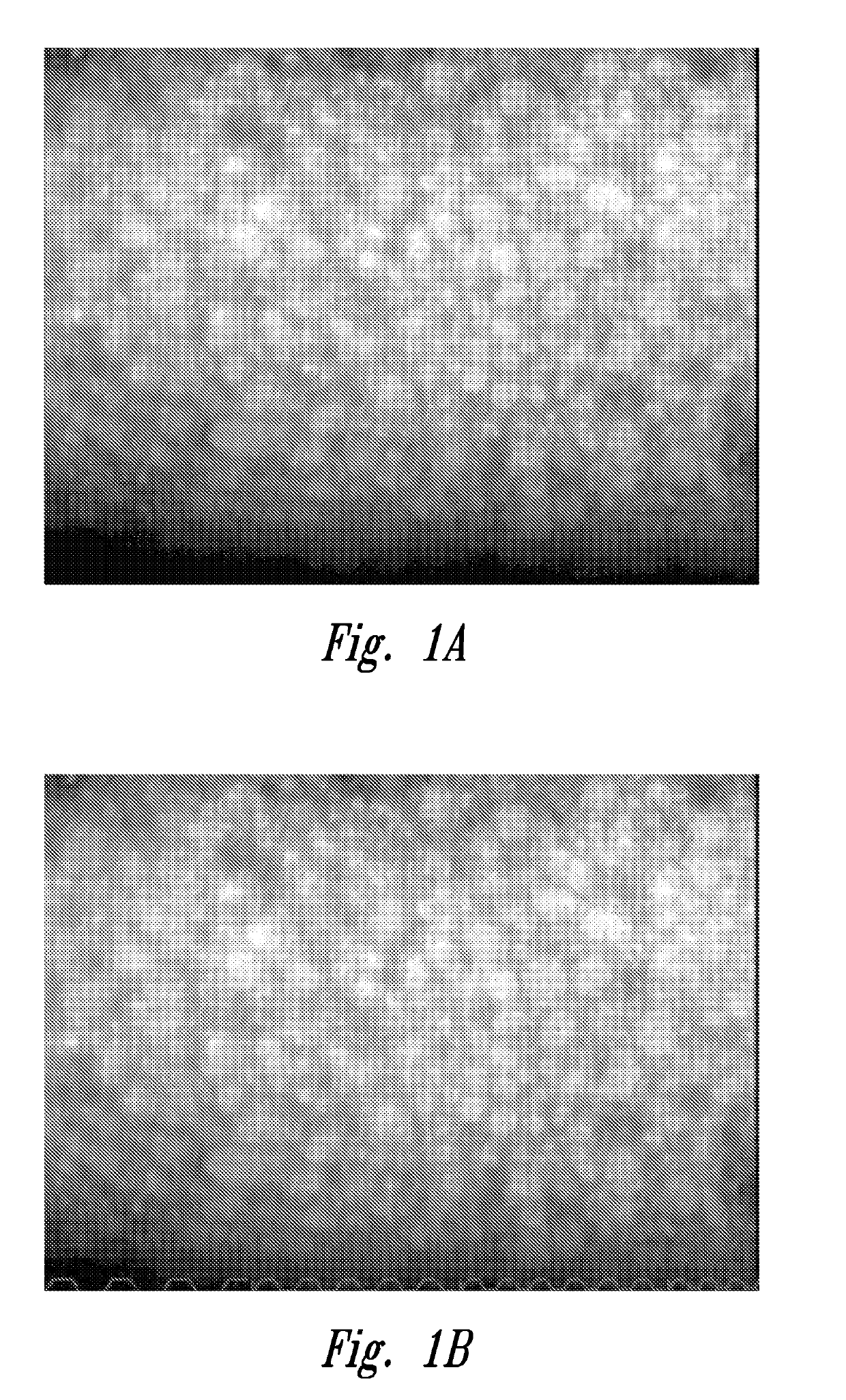
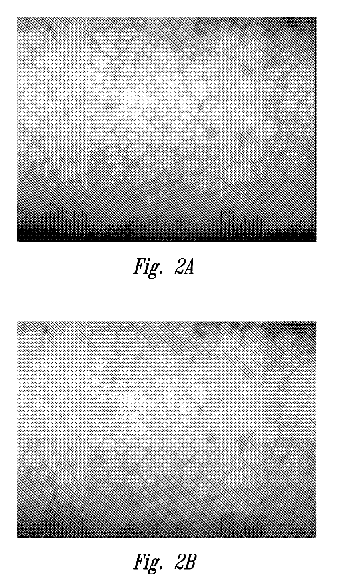
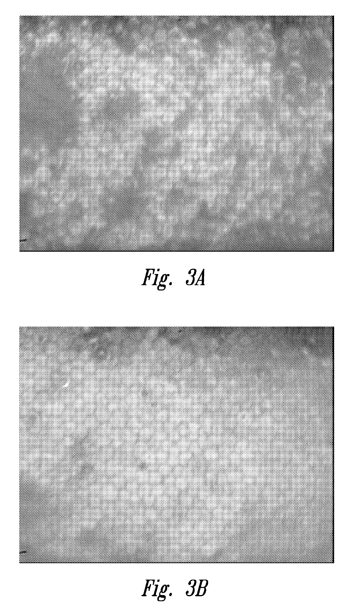
Re-Evaluation of Corneas at 34° C. Resulted in Identifying Corneas as Transplantable, Whereas the Same Corneas were Deemed not Suitable for Transplant (NSFT) Because of Poor Image Quality (Poor Endothelial Density) Upon Initial Evaluation of the Same Cornea at Room Temperature as in the Prior Art

[0081]As described above under “Background”, obtaining an accurate endothelial cell density (ECD) is a particularly important factor for quality assessment of harvested corneal tissue.

[0082]This example shows that at least a portion of poor image quality, poor endo (or dropout grade as shown in working Example 4 below) donor cornea endothelia, as initially judged at room temperature, achieved a state of better cell reflection and better cell border definition when evaluated at the cornea's natural functional temperature (e.g., 34° C.). While some corneas appeared to have endothelial dysfunction at room temperature, a portion of these exhibited evidence of cell viability equivalent to or bett...


PUM

PropertyMeasurementUnit
temperatureaaaaaaaaaa
temperaturesaaaaaaaaaa

Abstract

Provided are surprisingly effective methods for assessing corneal tissue quality, comprising: incubating corneal tissue in hypothermic corneal storage media to a temperature above ambient temperature; and assessing, using suitable microscopic examination, endothelial cell density and/or morphology and/or loss. The methods may additionally comprise; storing the corneal tissue in the hypothermic storage media at a temperature in the range of 2-8° C. prior to assessing the corneal tissue above ambient temperature. The methods may involve initially assessing corneal tissue quality at ambient temperature prior to assessing at a temperature above ambient temperature, with intermediate storage at 2-8° C.

Description

CROSS-REFERENCE TO RELATED APPLICATIONS[0001]This application claims the benefit of priority to U.S. Provisional Patent Application No. 62 / 347,034, filed 7 Jun. 2016 and entitled “METHOD FOR ASSESSING CORNEAL TISSUE QUALITY AND ENDOTHELIAL CELL DENSITY AND MORPHOLOGY”FIELD OF THE INVENTION[0002]Aspects of the present invention relate generally to methods for assessing corneal tissue quality, and more particularly to faster and more accurate methods for assessing measures of corneal tissue quality, including endothelial cell density and / or morphology as indicators of corneal tissue quality for purposes of improving outcomes with respect to corneal tissue storage and / or transplantation.BACKGROUND[0003]Effective corneal transplantation from harvested corneal tissue is dependent upon obtaining a good quality assessment of the harvested corneal tissue prior to transplantation. Such quality assessment is often performed multiple times prior to transplantation of the corneal tissue into a ...

Claims


Application Information

Patent Timeline
Patent Type & Authority Applications(United States)
IPC IPC(8): A01N1/02C12N5/079
CPCA01N1/0252A01N1/0221C12N5/0621G01N33/5005
Inventor FOX, ADAM M.HOOVER, CAROLINE
Owner CORNEAGEN INC
Why Eureka
Try Eureka