
Combined imaging detector for x-ray and nuclear imaging

a detector and nuclear imaging technology, applied in the field of combined imaging detectors, can solve the problems of insufficient clinical workflow between intervention (under c-arm control) and follow-up (e.g. by spectral imaging), and achieve the effects of less system sensitivity, easy reconstruction, and increased system sensitivity

Active Publication Date: 2019-08-08
KONINKLJIJKE PHILIPS NV
View PDF10 Cites 2 Cited by
  • Summary
  • Abstract
  • Description
  • Claims
  • Application Information

AI Technical Summary

Benefits of technology

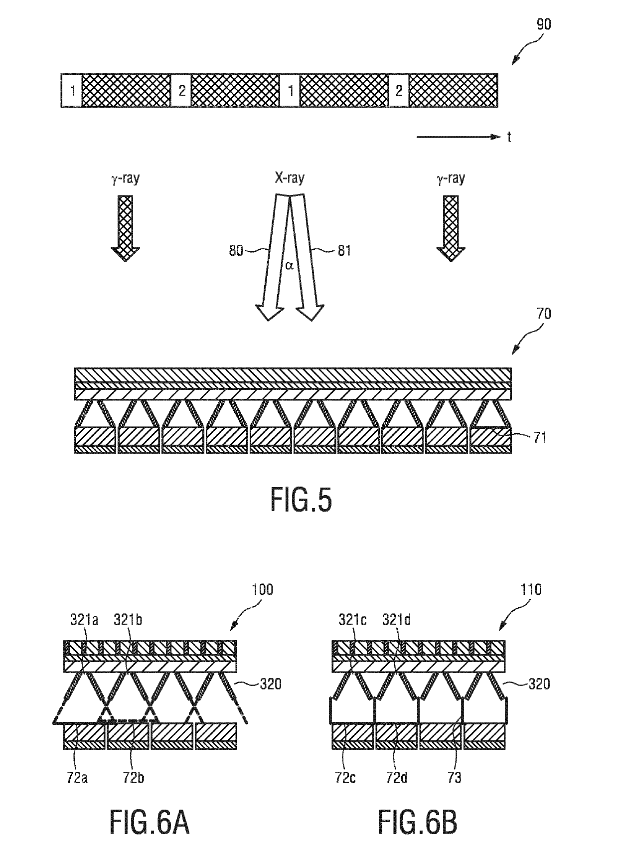
The patent describes a method to improve image-based intervention procedures by using both anatomical and molecular imaging techniques simultaneously. With this method, a better understanding of the anatomy and location of targeted areas can be achieved. This is useful for guiding surgeries and interventions that can track the location of tumors or other abnormal areas during needle insertion. The method involves using a special x-ray source that emits two split beams at different angles, which can help to reduce the impact of small missing X-ray areas on image quality.

Problems solved by technology

Especially the change between intervention (under C-arm control) and follow-up (e.g. by SPECT) is inadequate for the clinical workflow since the patient must be transported into another imaging system or another room and undergo multiple sessions including a liver-to-lung shunting evaluation (work-up) and then the actual therapy delivery.

Method used


Image

  • Combined imaging detector for x-ray and nuclear imaging
  • Combined imaging detector for x-ray and nuclear imaging
  • Combined imaging detector for x-ray and nuclear imaging

Examples


Embodiment Construction

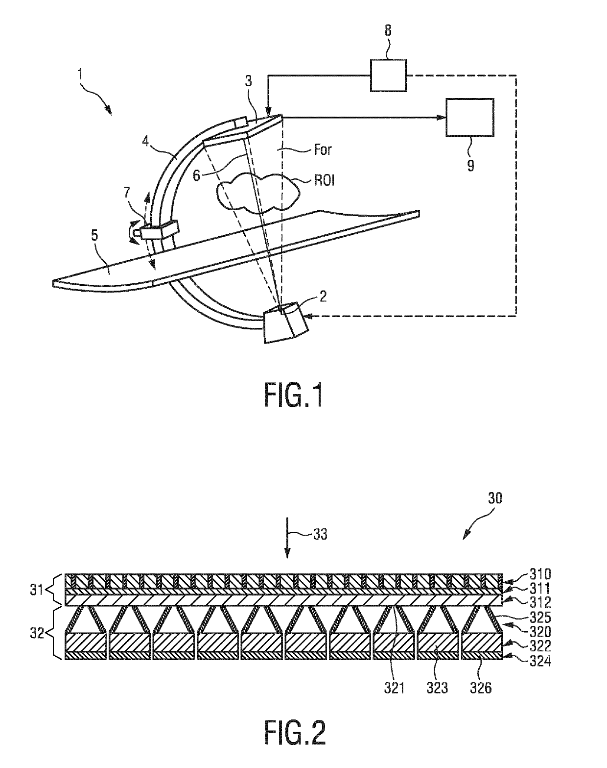
[0043]FIG. 1 illustrates an embodiment of an imaging system 1 according to the present invention. The imaging system 1 comprises an x-ray source 2, a combined imaging detector 3, a C-arm 4, and a patient table 5. The x-ray source 2 is attached to a first portion of C-arm 4 and the detector 3 is attached to a second portion of the C-arm 4. The x-ray source 2 and the detector 3 are so-positioned in order to measure x-ray transmission along path 6 between the x-ray source 2 and the detector 3. The field-of-view FOV of the x-ray source detector arrangement in FIG. 1 is illustrated by the short dashed lines that comprises path 6. The combined imaging detector 3 can be used for simultaneous (or subsequent or consecutive or alternate) detection of gamma and x-ray quanta to generate an x-ray image and a nuclear image for the same region of interest ROI.

[0044]The x-ray source 2 can be a standard x-ray source, although it is also contemplated to use a dual energy source in this position. Pref...


PUM


Abstract

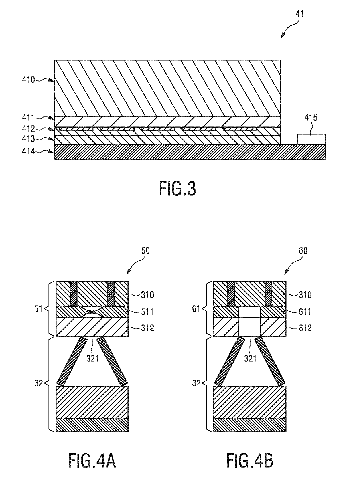
The invention relates to a combined imaging detector for detection of gamma and x-ray quanta comprising an x-ray detector (31) for generating x-ray detection signals in response to detected x-ray quanta and a gamma detector (32) for generating gamma detection signals in response to detected gamma quanta. The x-ray detector (31) and the gamma detector (32) are arranged in a stacked configuration along a radiation-receiving direction (33). The gamma detector (32) comprises a gamma collimator plate (320) comprising a plurality of pinholes (321), and a gamma conversion layer (322, 324) for converting detected gamma quanta into gamma detection signals.

Description

FIELD OF THE INVENTION[0001]The invention relates to a combined imaging detector for detection of gamma and x-ray quanta and to a corresponding imaging system. It finds application in the medical field, more particularly in the fields of medical imaging and medical interventions, and may be used for example to provide an x-ray image and a corresponding nuclear image of a region of interest. A gamma detector is also disclosed.BACKGROUND OF THE INVENTION[0002]In various medical imaging procedures it is beneficial to provide both an x-ray image and a nuclear image of a region of interest. The x-ray image typically provides structural information indicative of the anatomy of the region of interest. The nuclear image, defined herein to mean an image indicative of radiotracer distribution in an object, is generated based on detected gamma quanta. The nuclear image may for example be a gamma scintigraphy or a SPECT image and typically provides functional, or physiological information relat...

Claims


Application Information

Patent Timeline
Patent Type & Authority Applications(United States)
IPC IPC(8): G01T1/164A61B6/06A61B6/00
CPCG01T1/1644G01T1/1648G01T1/1647A61B6/06A61B6/4258A61B6/4291A61B6/037A61B6/4233A61B6/4417A61B6/4441A61B6/486A61B6/487A61B6/5235G01T1/1611
Inventor WIECZOREK, HERFRIED KARLJACOBS, JOHANNES WILHELMUS MARIASTEGEHUIS, HERMANRADAELLI, ALESSANDROKOK, CHRISTIAANALVING, PETER LEX
Owner KONINKLJIJKE PHILIPS NV
Learn More