
Cancer modeling platforms and methods of using the same

a technology of cancer modeling and platform, applied in the field of in vitro cell constructs, can solve the problem of difficult choice of absolute best drug for a particular patien

Pending Publication Date: 2019-06-20
WAKE FOREST UNIV HEALTH SCI INC
View PDF1 Cites 9 Cited by
  • Summary
  • Abstract
  • Description
  • Claims
  • Application Information

AI Technical Summary

Benefits of technology

The invention is a method for creating tumor models using live tumor cells and live benign cells. These cells can be obtained from a subject and combined to form an organoid, which can be used for evaluating tumor cell behavior and response to drugs or other compounds. A device is also described, which can contain multiple organoids and can be used to evaluate tumor cell growth and migration. Overall, the invention provides a way to better understand the biology of tumor cells and develop new treatments for cancer.

Problems solved by technology

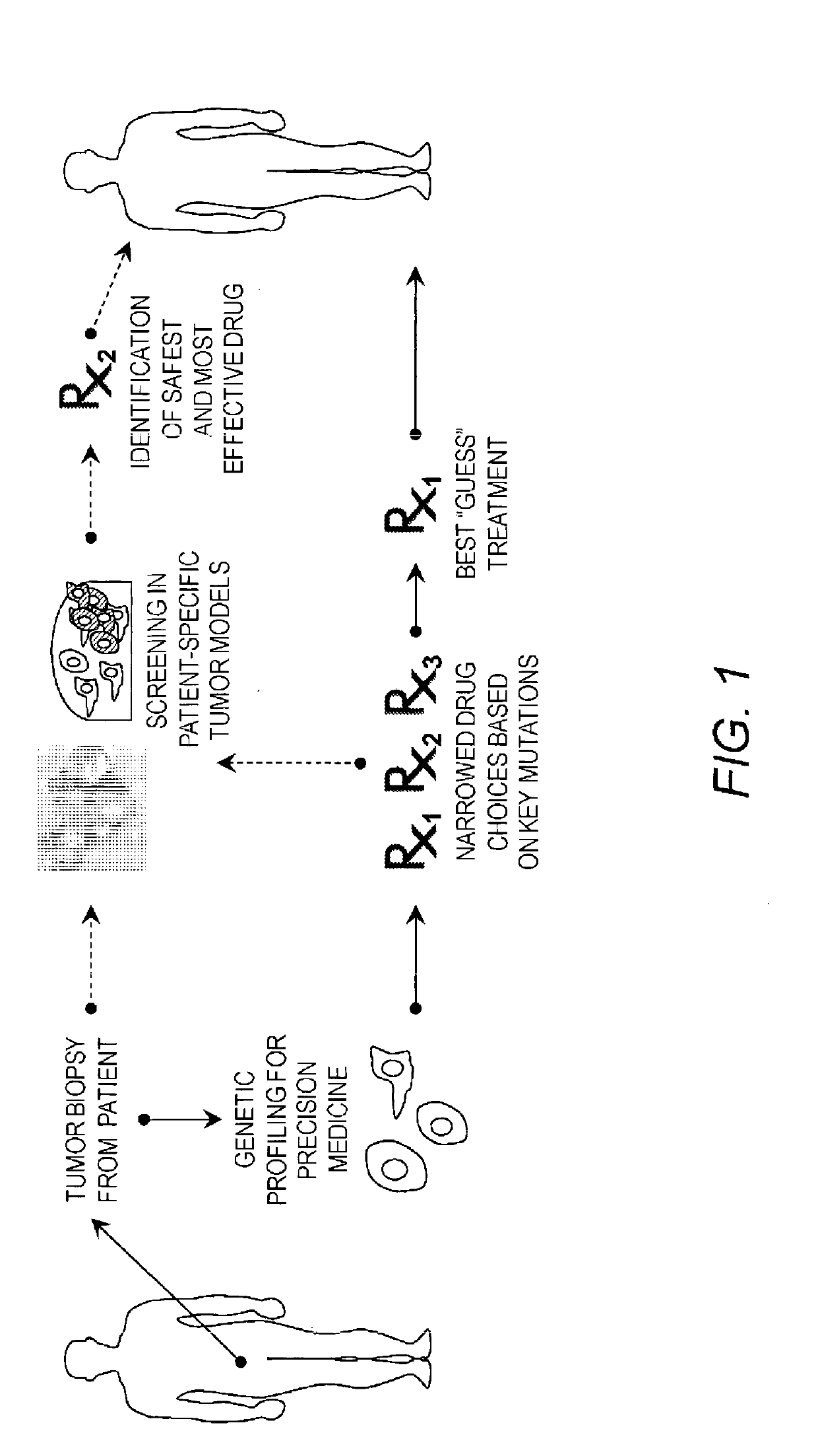
Currently, genetic profiling can be performed on tumor biopsies to narrow down drug choices based on key mutations, but choosing the absolute best drug for a particular patient remains difficult.

Method used


Image

  • Cancer modeling platforms and methods of using the same
  • Cancer modeling platforms and methods of using the same
  • Cancer modeling platforms and methods of using the same

Examples


example 1

[0081]The first objective of this project was to evaluate a 3D model of a colorectal cancer tumor surrounded by normal host tissue. The engineered concentric ring construct consists of a colorectal tumor core and a colon epithelial outer ring, to model a tumor in a reductionist, yet physiologically relevant environment. This construct was used to measure the growth of the tumor core, any observed invasion of the tumor cells into the epithelial layer, and the efficacy of drugs. The constructs were created with cells suspended in a hyaluronic acid and gelatin-based hydrogel. The tumor cores were made using one of four colorectal cancer cells types, thereby providing models of 4 levels of malignancy. The HCT116 cells were the most malignant, followed by the HT29, SW480, and Caco2 cells. Caco2 cells were also employed in the outer ring, as they best resemble healthy epithelial cells. The tumor core was fluorescently labeled and fluorescent imaging was used to monitor the tumor core over...

example 2

[0084]3D tumor organoids were microengineered directly from fresh tumor biopsies to provide a patient-specific model system with which treatment optimization can be performed before initiation of therapy. Here, the initial implementation of this platform is demonstrated using a mesothelioma tumor biospecimen removed from a patient. Shown is the ability to biofabricate and maintain viable 3D tumor constructs within a tumor-on-a-chip microfluidic device. Second, it is demonstrated that on-chip chemotherapy screening mimics patient response to the drugs cisplatin and pemetrexed. Finally, it is demonstrated in vitro the effectiveness of a candidate compound identified through genetic biomarker assessment.

Methods

Thin Film Microfluidic Device Fabrication

[0085]The microfluidic device fabrication approach is based on methods reported elsewhere. Channels were formed in double-sided adhesive film using a cutting plotter (GraphTec-CE6000-40) and the bottom surface of the patterned layer was ad...

example 3

[0110]Multiple sets of patient tumor-derived organoids have been generated with over a 90% take rate in vitro (versus below 10% in 2D cell cultures), which is favorably compared to a reported 25-33% take rate in patient-derived xenograft (PDX) models where only the most aggressive biospecimens prosper. This poor take rate limits the application of PDX technology to predictive diagnostics for most cancer patients. Our tumor organoid systems have subsequently been employed in a drug screens to demonstrate that like patients, their tumor organoids maintain selective response to different drugs. To date sets of patient-specific organoids have been created from a diverse group of primaries and their metastatic sites including colorectal cancer, high and low grade appendiceal metastatic sites to omentum ovary and liver, peritoneal mesothelioma, and extremity sarcoma. Described herein are several examples, demonstrating 1) correlation between tumor organoid drug response and the drug respo...


PUM


Abstract

Described herein are in vitro cell constructs (or “organoids”) useful as a tumor model, the constructs comprising live tumor cells from a subject. In some embodiments, provided are in vitro cell constructs comprising: (a) a core comprised of live tumor cells; and (b) a shell surrounding (e.g., encapsulating) the core, the shell comprised of live benign cells (e.g., tissue cells, benign or differentiated tumor cells, etc.). Also described herein are methods of making and using such constructs. Further provided are devices useful for evaluating tumor cells in vitro, comprising: (a) a microfluidic device having a chamber, and a channel in fluid communication with the chamber; (b) a live tumor cell construct (e.g., an organoid) in the chamber; (c) a growth media in the chamber and the channel; (d) a pump operatively associated with the chamber and channel and configured for circulating the media from the chamber through the channel and back to the chamber; (e) a microporous membrane (e.g., a TRANSWELL® microporous membrane) in the channel and positioned so that the media flows therethrough.

Description

RELATED APPLICATION INFORMATION[0001]This application claims the benefit of U.S. Provisional Patent Application Ser. No. 62 / 370,320, filed Aug. 3, 2016, and U.S. Provisional Patent Application Ser. No. 62 / 522,313, filed Jun. 20, 2017, the disclosure of each of which are incorporated herein by reference in their entirety.STATEMENT OF GOVERNMENT SUPPORT[0002]This invention was made with government support under grant numbers 1U54TR001362-01 and 5UL1TR001420-03 awarded by the National Institutes of Health. The government has certain rights in this invention.FIELD OF THE INVENTION[0003]This application relates to in vitro cell constructs (or “organoids”) that may be used as a tumor model along with methods for making and using the same.BACKGROUND[0004]Precision medicine shows incredible promise for improving treatment in cancer patients. Currently, genetic profiling can be performed on tumor biopsies to narrow down drug choices based on key mutations, but choosing the absolute best drug...

Claims


Application Information

Patent Timeline
IPC IPC(8): G01N33/50C12M1/12C12M3/06C12M1/26C12M1/42C12M1/34
CPCG01N33/5011C12M25/16C12M23/16C12M33/14C12M35/08C12M41/46G01N2500/10C12N5/0012C12N5/0679C12N5/0693C12N2503/02C12N2503/04C12N2513/00C12N2533/30C12N2533/80G01N33/5008C12N5/0634G01N33/5088C12M29/04
Inventor SKARDAL, ALEKSANDERVOTANOPOULOS, KONSTANTINOS
Owner WAKE FOREST UNIV HEALTH SCI INC
Learn More