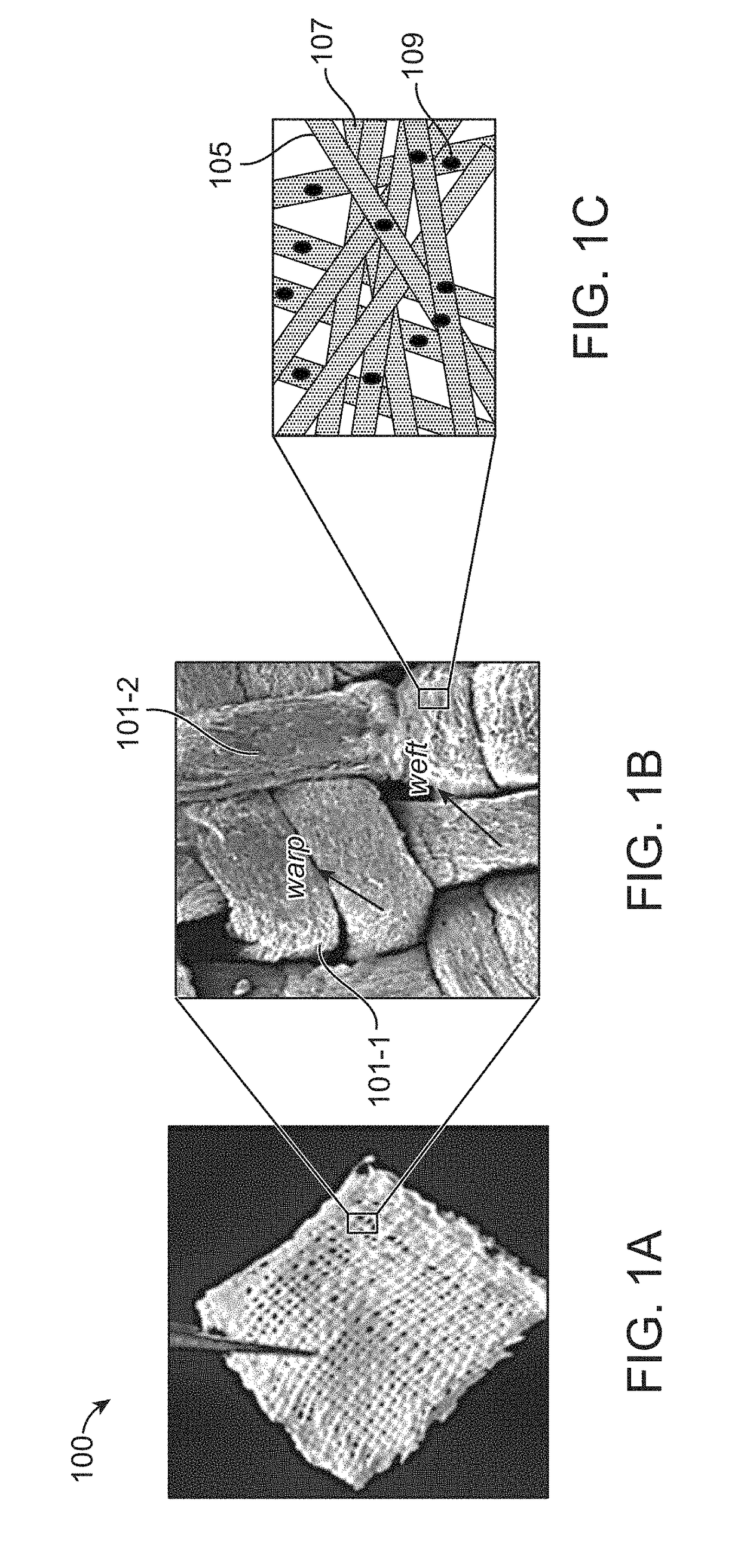
Suturable woven implants from electrospun yarns for sustained drug release in body cavities
a technology of woven implants and electrospun yarns, which is applied in the direction of prosthesis, surgery, other medical devices, etc., can solve the problems of not having any marketed products or clinically applicable devices with long-term effectiveness, and achieve the effect of controlled and sustained release of therapeutic agents
- Summary
- Abstract
- Description
- Claims
- Application Information
AI Technical Summary
Benefits of technology
Problems solved by technology
Method used
Image
Examples
example 1
on of Woven Fabric from Drug-Laden 1D Electrospun Single Polymeric Yarns
[0064]A drug-laden medical fabric was developed for implantation by feeding paclitaxel-loaded, one-dimensional uniform and continuous electrospun polymeric yarns with continuous fibers into a weaving loom and a conventional method of plain weaving was adopted for weaving. Woven fabrics were fabricated by interlacing two sets of drug-loaded yarns as warps and wefts perpendicular to each other on a computer-controlled table top plain weaving loom.
[0065]The drug-laden yarns were obtained by electrospinning a polydioxanone (PDS) containing organic solution with 1 to 50 w / w % of drug (paclitaxel) at optimized conditions of flow-rate in the range of 1 to 5 ml / h, voltage 10 to 15 kV, collector rotation 500 to 1000 rpm and yarn-uptake rate of 0.25 to 0.75 m / min to obtain 1D continuous yarn with high encapsulation rate of 85 to 95% loaded with amorphous drug within the polymeric matrix. Similarly, other biodegradable sin...
example 2
cterization of the Drug-Laden Woven Fabric
[0068]The fabrics from Example 1 was scanned with Scanning Electron Microscope (SEM) and the average diameters of the electrospun polymeric yarns were measured with and without the drug loaded onto them.
[0069]FIGS. 2A-2D show SEM images of electrospun polymeric yarns. FIG. 2A shows the SEM images of electrospun PDS polymeric yarns. It shows smooth, continuous, nano fibrous yarns with an average diameter of 250-280 μm (3-0 sutures USP dimension). The electrospun polymeric yarns with individual fibers having an average diameter of 450 (400-500 nm range) nm are shown in FIG. 2B. FIG. 2C shows the SEM image of 10 w / w % PTX-loaded PDS polymeric yarn having an average diameter of 400-500 nm. FIG. 2D shows SEM image of PTX-loaded PLLA polymeric yarns. PTX inclusion within the PDS yarns did not alter its morphology. Similarly, non-woven electrospun mats with average fiber diameter of ˜750 nm were fabricated by conventional electrospinning for compar...
example 3
ing for Encapsulation of the Drug within the Yarn
[0071]The encapsulation of drug (paclitaxel) within the polymeric matrix of the fabrics from Example 1 was tested using Fourier-Transform Infrared spectroscopy (FTIR) technique. FIG. 4 shows the FTIR spectra of the composite yarns revealing the encapsulation of paclitaxel within the polymeric matrix of PDS (A) and PLLA (B). The retention of CN stretching peak of the drug at 1251 cm−1 was measured to confirm drug entrapment in the polymeric yarns. The peaks at 1128 cm−1 and 1732 cm−1 indicated the ether backbone of PDS polymer and carbonyl groups of both PTX and PDS. The presence of ether-ester backbone of PDS confers greater flexibility as well as elongation to polymeric matrix.
PUM
| Property | Measurement | Unit |
|---|---|---|
| Time | aaaaa | aaaaa |
| Diameter | aaaaa | aaaaa |
| Diameter | aaaaa | aaaaa |
Abstract
Description
Claims
Application Information
 - R&D
- Intellectual Property
- Life Sciences
- Materials
- Tech Scout
- Unparalleled Data Quality
- Higher Quality Content
- 60% Fewer Hallucinations
Browse by: Latest US Patents, China's latest patents, Technical Efficacy Thesaurus, Application Domain, Technology Topic, Popular Technical Reports.
© 2025 PatSnap. All rights reserved.Legal|Privacy policy|Modern Slavery Act Transparency Statement|Sitemap|About US| Contact US: help@patsnap.com


