
Methods of Mapping Protein Variants

a protein variant and variant technology, applied in the field of protein variant analysis, can solve the problems of inconvenient pre-clinical or clinical pk study of biopharmaceuticals, limited approach, and decrease in the amount of recombinant protein of interes

Inactive Publication Date: 2019-01-24
HEXAL AG
View PDF0 Cites 5 Cited by
  • Summary
  • Abstract
  • Description
  • Claims
  • Application Information

AI Technical Summary

Benefits of technology

The present invention relates to a method for analyzing protein variants of a recombinant protein of interest in liquid samples of a mammal. The method involves immobilizing the recombinant protein on a solid support, eluting it from the support, and analyzing the protein variants using an analytical separating method such as HPLC, CE, or MS. The method can be used to analyze pharmacokinetic parameters of the protein variants, including deamidation, oxidation, N-terminal and C-terminal heterogeneity, isomerization, glycation, and disulfide isoforms. The method can also be used to analyze one or more pharmacokinetic parameters of the protein variants, such as glycosylation variants. The method is particularly useful for analyzing antibodies and fusion proteins. The invention also provides a solid support for immobilizing the recombinant protein and an internal standard for analysis. The method can be used with liquid samples prepared in a multi-well plate. The liquid samples can be body fluids such as serum, plasma, urine, cerebral spinal fluid, amniotic fluid, saliva, sweat, ejaculate, tears, phlegm, vaginal secretion, vaginal wash, and colonic wash.

Problems solved by technology

Further, the amount of the recombinant protein of interest declines over time.
However, this approach has limitations because of the assumption that the only change in the molecule during enrichment is the glycan structure.
Glycan heterogeneity can also lead to some ambiguity of the results.
Thus, this approach is not suitable for pre-clinical or clinical pK studies of biopharmaceuticals, such as therapeutic recombinant antibodies or fusion proteins.
However, the assay allowed analysis only up to 14 days post-administration in a rather high sample volume and is prone to contamination with other serum proteins.
Also, this method is subject to interference from glycans released from other serum proteins or other glycans of the same molecule for glycoproteins with more than one glycosylation sites.
However, this method requires a sample volume of 0.5 ml, which often may not be available, e.g., in preclinical studies using rodents.
However, analysis on modifications other than glycosylation was not performed.
However, this approach has limitations because of the assumption that the only change in the molecule during enrichment is the charge form.
Thus, this approach is not suitable for pre-clinical or clinical pK studies of biopharmaceuticals, such as therapeutic recombinant antibodies or fusion proteins.
However, this method requires a sample volume of 0.5 ml, which often may not be available, e.g., in preclinical studies using rodents.

Method used


Image

  • Methods of Mapping Protein Variants
  • Methods of Mapping Protein Variants
  • Methods of Mapping Protein Variants

Examples


example 1

ing of Disulfide Isoforms of an IgG1 Antibody

Preclinical Rabbit Study

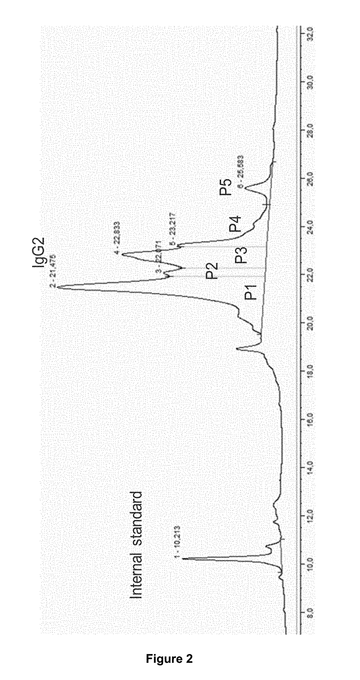
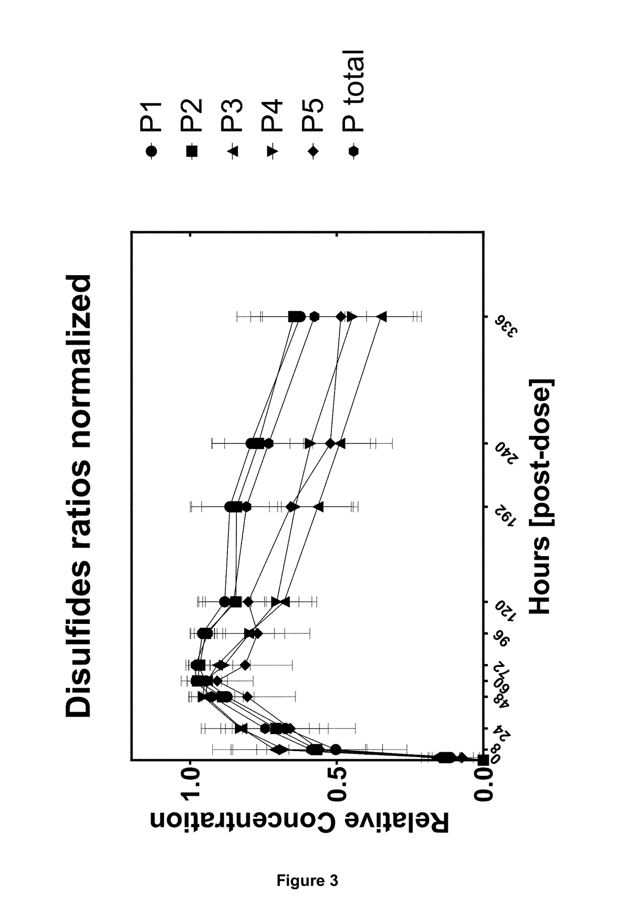
[0134]The preclinical study was performed in cynomolgus monkeys. Following single subcutaneous administration of 3 mg kg-1 body weight of an IgG2 mAb1, blood samples were drawn over a period of time including one pre-dose blood sample. Serum samples were taken at 10 time points at 8, 24, 48, 60, 72, 96, 120, 192, 240 and 336 hours post administration. Concentration of mAb1 in serum was determined by ELISA. From remaining serum 2×50 μl aliquots were used for PK profiling. The first aliquot was analyzed and the second aliquot served as back-up aliquot.

Buffer Preparation

[0135]Acidification solution, 1 mM hydrochloric acid pH 3.0: 100 μl of 1 N (1 M) hydrochloric acid were pipetted into a 100 ml bottle filled with 99.8 g ultra-pure water. After briefly mixing the pH was measured and should be 3.0. The solution has to be prepared fresh before use.

[0136]Blocking buffer, 0.5 M ethanolamine, 0.5 M NaCl pH 8.3: 2.92 g NaCl ...

example 2

ing of N-Terminal Leader Peptide Extensions on IgG1 Light Chains

[0153]Antibodies can contain variations at their N-termini of both heavy and light chain. These extensions can result from incomplete processing during protein biosynthesis. The extension can for example contain charged or hydrophobic amino acids that can influence the physico-chemical properties (e.g. binding of the antigen). In the present case a portion of the IgG1 biopharmaceutical has an N-terminal leader peptide extension on one light chain. The affinity purification work-flow is illustrated in FIG. 5.

Preclinical Rabbit Study

[0154]The preclinical study was performed in Himalayan rabbits following intraveneous administration of 15 mg / kg body weight over 30 minutes of a humanized IgG1 mAb2 antibody. The mAb2 antibody contains to some extend an N-terminal leader peptide extension on the light chain. The blood samples were drawn over a period of time including one pre-dose blood sample. Sampling was performed 10, 20 a...


PUM

PropertyMeasurementUnit
volumeaaaaaaaaaa
molecular massaaaaaaaaaa
volumeaaaaaaaaaa

Abstract

The present invention relates to a method for analysing protein variants of a recombinant protein of interest, such as antibodies or Fc-fusion proteins, in a liquid sample of a mammal. Specifically the method comprises a step of affinity purifying the recombinant protein of interest from the sample together with an internal standard, and analyzing the protein variants using an analytic separating method such as HPLC, capillary electrophoresis or MS. This method is particularly suited to measure pharmacokinetic parameters of a recombinant protein of interest, such as a biopharmaceutical, in a mammal in clinical or pre-clinical studies. It allows for the use of a small sample volume and the possibility to operate with high throughput, such as in a 96-well plate sample preparation. It also provides high sensitivity and allows analysis of protein variants individually.

Description

FIELD OF THE INVENTION[0001]The present invention relates to a method for analysing protein variants of a recombinant protein of interest, such as antibodies or Fc-fusion proteins, in a liquid sample of a mammal. Specifically the method comprises a step of affinity purifying the recombinant protein of interest from the sample together with an internal standard, and analyzing the protein variants using an analytic separating method such as HPLC, capillary electrophoresis or MS. This method is particularly suited to measure pharmacokinetic parameters of a recombinant protein of interest, such as a biopharmaceutical, in a mammal in clinical or pre-clinical studies. It allows for the use of a small sample volume and the possibility to operate with high throughput, such as in a 96-well plate sample preparation. It also provides high sensitivity and allows analysis of protein variants individually.BACKGROUND OF THE INVENTION[0002]Biological drugs belong to the most complex pharmaceuticals...

Claims


Application Information

Patent Timeline
Patent Type & Authority Applications(United States)
IPC IPC(8): G01N33/68G01N33/543
CPCG01N33/6848G01N33/6842G01N33/54326G01N33/6818G01N2440/16G01N2440/20G01N2570/00G01N33/6803G01N2440/00G01N2440/38
Inventor HIGEL, FABIANSEIDL, ANDREAS
Owner HEXAL AG
Learn More