
Quantitative dermoscopic melanoma screening

a dermoscopic and melanoma technology, applied in the field of skin disease detection, can solve the problems of difficult decision on which lesions to biopsy, immense human and financial costs, and the most lethal skin cancer, and achieve the effect of greater sensitivity, specificity and overall diagnostic accuracy

Active Publication Date: 2018-08-23
THE ROCKEFELLER UNIV
View PDF0 Cites 5 Cited by
  • Summary
  • Abstract
  • Description
  • Claims
  • Application Information

AI Technical Summary

Benefits of technology

This patent is about improving the evaluation of skin lesions through the use of algorithms that can detect and analyze image data from reflected or fluoresced light. The goal is to increase sensitivity, specificity, and overall diagnostic accuracy, and to provide quantitative imaging biomarkers that can aid in the diagnosis of skin cancer and other abnormal skin conditions. Additionally, the patent combines a dermatoscope, digital camera, and automated screening by computer vision to bridge the gap between invasive and noninvasive pathological analyses.

Problems solved by technology

Melanoma, the most lethal skin cancer, incurs immense human and financial cost.
After invasive biopsy or excision, diagnosis obtained by histopathologic evaluation is nearly 100% accurate; however, deciding which lesions to biopsy is challenging.

Method used


Image

  • Quantitative dermoscopic melanoma screening
  • Quantitative dermoscopic melanoma screening
  • Quantitative dermoscopic melanoma screening

Examples


Embodiment Construction

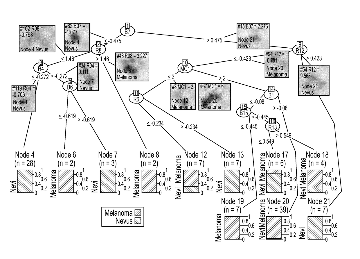
[0016]One embodiment of the present invention is directed to a system including a camera, a mechanical fixture for illuminating the subject's skin and positioning the camera fixedly against the subject's skin, at least one processor adapted to perform the clock sweep algorithm, and at least one output device to display diagnostic information.

[0017]The camera is preferably a digital camera and may include a charged coupled device (CCD) sensor or complementary metal oxide semiconductor (CMOS), as known in the art. The camera may be a commercially available portable camera with an integrated illumination system or flash and a sensor array detecting Red Green and Blue (RGB) light. Alternatively an external illumination system may be provided and the camera sensor array may be adapted to receive “hyperspectral” light, meaning light divided into more spectral wavelength bands than the conventional RGB bands, which may be in both the visible and non-visible range. Details concerning operab...


PUM


Abstract

A standardized, quantitative risk assessment method and apparatus for noninvasive melanoma screening. The apparatus and methods generate a melanoma Q-Score which calculates and displays a probability that a skin lesion is melanoma.

Description

[0001]This application claims the benefit of U.S. Provisional Patent Application No. 62 / 204,763, filed Aug. 13, 2015, which is incorporated by reference in its entirety.BACKGROUND OF THE INVENTION[0002]The invention is directed to detection of skin disease from image analysis of a skin lesion.[0003]Melanoma, the most lethal skin cancer, incurs immense human and financial cost. Early detection is critical to prevent metastasis by removal of primary tumors. The early lateral growth phase is a vastly preferable detection window to the subsequent phase of metastatic initiation. Optical detection technologies for automated quantitative metrics of malignancy are needed to more accurately guide decisions regarding the need to biopsy and to make preoperative determination of adequate margins for surgical excision. After invasive biopsy or excision, diagnosis obtained by histopathologic evaluation is nearly 100% accurate; however, deciding which lesions to biopsy is challenging. Only 3% to 2...

Claims


Application Information

Patent Timeline
Patent Type & Authority Applications(United States)
IPC IPC(8): A61B5/00G06T7/00G06T7/68G06K9/62
CPCA61B5/444A61B5/0071A61B5/0077A61B5/743G06T7/0012G06T7/68G06K9/628G06T2207/10024G06T2207/10064G06T2207/20081G06T2207/30088G06T2207/30096A61B5/0075A61B5/7275G06T2200/36G16H50/30G16H50/20G16H30/40G16H30/20G06F18/2431
Inventor GAREAU, DANIEL
Owner THE ROCKEFELLER UNIV
Learn More