
Microscope or endoscope assembly and method for reducing specular reflections

a technology of endoscope and endoscope, which is applied in the field of microscope or endoscope assembly, can solve the problems of purkinje reflex, strong lightening artifact, and other surgical microscope problems, and achieve the effect of reducing the number of spherical reflections

Inactive Publication Date: 2018-06-21
LEICA INSTR SINGAPORE PTE
View PDF0 Cites 0 Cited by
  • Summary
  • Abstract
  • Description
  • Claims
  • Application Information

AI Technical Summary

Benefits of technology

The patent is about a microscope or endoscope assembly that uses a polarization rotor to adjust the angle of polarization in the light that passes through it. This allows for better distinction between incident and reflected light, and can result in a more compact assembly. The use of a quarter-wave plate as a polarization rotor can also help to maintain information about the three-dimensional structure of tissue layers. By adding a further polarizing beam splitter in the illumination or observation path, the efficiency of illumination can be further increased.

Problems solved by technology

However, the illumination used for the red reflex causes strong lightening artifacts called Purkinje reflexes, which are essentially specular reflections.
The same problem also exists in other surgical microscopes which are used for other applications.
If the tissue is illuminated, specular reflections may be generated which deteriorate the image quality.

Method used


Image

  • Microscope or endoscope assembly and method for reducing specular reflections
  • Microscope or endoscope assembly and method for reducing specular reflections
  • Microscope or endoscope assembly and method for reducing specular reflections

Examples


Embodiment Construction

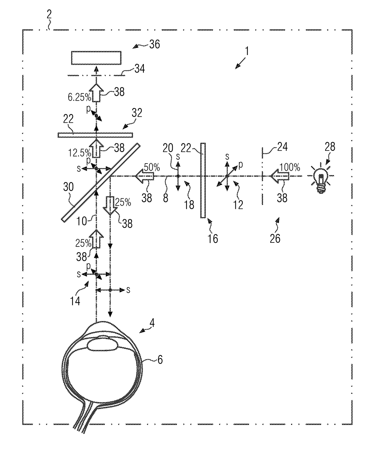
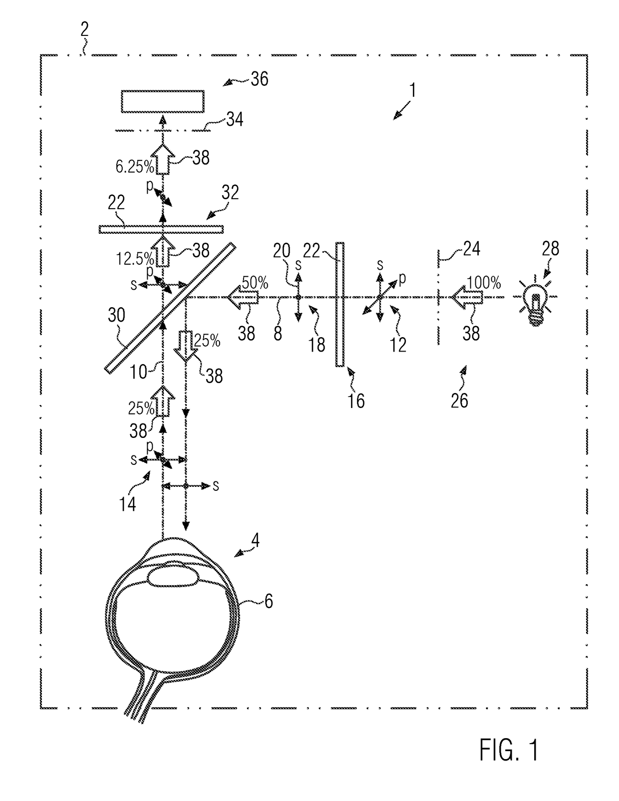
[0047]First, the configuration of a microscope or endoscope assembly 1 according to the invention will be explained with reference to FIG. 1.

[0048]The microscope and endoscope assembly 1, in short “microscope assembly” in the following, is adapted to be part of or to be installed in a surgical microscope or endoscope 2, in particular an ophthalmic microscope, without being limited to such an application. The microscope or endoscope 2 is shown only schematically and only in FIG. 1.

[0049]It is further shown to comprise an observation region 4, in which an object 6 to be observed is placed in operation of the microscope or endoscope 2. The observation region 4 may be part of the microscope or endoscope assembly 1. Although the observation region 4 may not be a structural part of the microscope assembly 1 or the microscope or endoscope 2, it is nonetheless a functional part at least in operation, as the technical effects of the components of the microscope assembly 1 or the microscope o...


PUM


Abstract

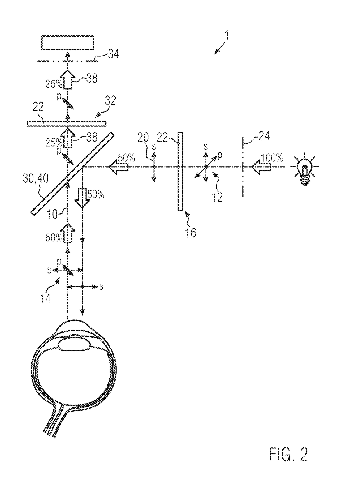
The invention relates to a microscope or endoscope assembly (1), in particular for a surgical microscope such as an ophthalmic microscope. Especially in eye surgery, but also in other application concerning life tissue, specular reflections of the illumination light are unwanted because they may hide important information contained in a diffuse reflection at the same location. In order to suppress such unwanted specular reflection, the microscope or endoscope assembly according to the invention comprises an observation region (4), in which an object (6) to be observed, such as an eye, can be arranged. The assembly further comprises an illumination light path (8) which extends from an illumination light entry region (24), where illumination light enters into the assembly, to the observation region. Further, an observation light path (10) is comprised, which extends from the observation region (4) to a light exit region (34), where the light leaves the assembly and may be collected by an observation subassembly (36), such as at least one camera and / or at least one ocular. In the illumination light path, a first polarizing subassembly (16) is arranged, to create polarized illumination light (18). In the observation light path (10) a second polarizing subassembly (32) is arranged, which is configured to filter out the polarized illumination light (18) passing the first polarizing subassembly (16). To ensure coaxial illumination and observation of the object, a beam splitter (30, 40) is provided, which is arranged in the illumination light path (8) between the light entry region (24) and the observation region (4) and in the observation light path (10) between the observation region (4) and the light exit region (34). By the complementary filtering action of the first and second polarizing subassembly, specular reflections by the object can be suppressed and / or eliminated.

Description

CROSS-REFERENCE TO RELATED APPLICATIONS[0001]The present application claims priority of European Patent Application No. 16204933.2 filed Dec. 19, 2016, the entire disclosure of which is incorporated herein by reference.FIELD OF THE INVENTION[0002]The invention relates to a microscope or an endoscope assembly, in particular for a surgical microscope such as an ophthalmic microscope. The invention further relates to a retrofit kit for generating such a microscope assembly and to the use of such a microscope assembly as well as to a method for operating a microscope, in particular a surgical microscope.[0003]Ophthalmic microscopes are used for operations on the eye, such as cataract surgery. In cataract surgery, ophthalmologists rely on the red reflex to see the capsule, lens and anterior chamber structure of the eye. The red reflex is, in essence, a total light reflection within the eye, which can be achieved when the eye is illuminated from the right angle and from the right distance...

Claims


Application Information

Patent Timeline
IPC IPC(8): G02B21/06A61B1/00A61B3/13A61B90/20G02B27/28G02B27/14G02B21/00G02B23/24
CPCG02B21/06A61B1/00186A61B3/13A61B90/20G02B27/286G02B27/14G02B21/0012G02B23/2461G02B21/0028G02B21/0032G02B21/0068G02B21/0092G02B23/2407A61B3/158A61B3/125
Inventor THEMELIS, GEORGE
Owner LEICA INSTR SINGAPORE PTE
Learn More