
Determination of a clinical characteristic using a combination of different recording modalities

a clinical characteristic and recording modality technology, applied in the field of determining the clinical characteristic of a body vessel segment, can solve the problems of inaccurate geometric representation of the stenosis geometry, low spatial resolution, and estimation of blood flow using the vascular cross-section, so as to achieve the effect of less easily obtained or extracted or determinable, and less radiation dose, and simple determination of clinical characteristics

Active Publication Date: 2018-03-01
SIEMENS HEALTHCARE GMBH
View PDF9 Cites 2 Cited by
  • Summary
  • Abstract
  • Description
  • Claims
  • Application Information

AI Technical Summary

Benefits of technology

This patent describes a method for analyzing blood vessels using angiography and creating a 3D model of the vessels using computed tomography (CT) scans. The method takes advantage of the fact that only the part of the vessel where the blockage is located needs to be analyzed, instead of the entire vessel. This reduces the amount of radiation that patients are exposed to and ensures accurate results. The method uses global features to analyze the entire tree of blood vessels, while also using local features to provide specific information about blockages. The method also checks the accuracy of the results by comparing them to a global feature. Overall, this method improves the accuracy and efficiency of analyzing blood vessels and their treatment plan.

Problems solved by technology

A disadvantage of this is the comparatively low spatial resolution and thus an inaccurate geometric representation of the stenosis geometry.
A disadvantage of this is the estimation of the blood flow using the vascular cross-sections.
Even small errors here may have major consequences.
Estimating the blood flow using contrast medium dynamics in the angio-FFR method is also laborious and difficult.
Another disadvantage is that the angio-FFR method does not provide any information on a status of the myocardial mass, which is important, for example, in order to be able to detect any pre-existing damage and to take account of the information during treatment.
Geometric information about the entire vascular tree may only be obtained with great difficulty, in part due to the relatively small detectors normally used in angiography.

Method used


Image

  • Determination of a clinical characteristic using a combination of different recording modalities
  • Determination of a clinical characteristic using a combination of different recording modalities

Examples


Embodiment Construction

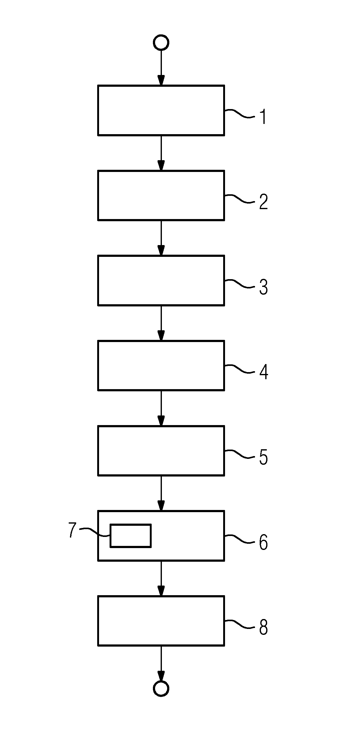
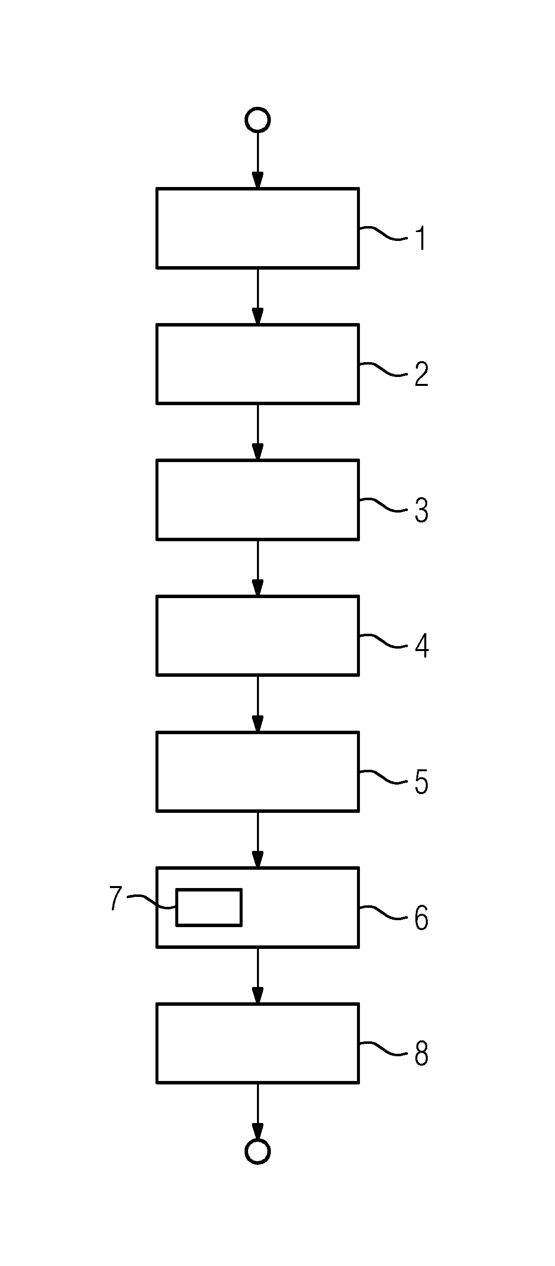
[0030]In the method represented in FIG. 1, a computing device identifies (e.g., is provided) a three-dimensional reconstruction of a body vessel (e.g., a coronary artery) in a first act. The body vessel includes, for example, a complete body vascular tree of the coronary artery and also has a body vessel segment (e.g., a vascular branch). In the example shown, the body vessel segment is a body vessel segment of the coronary artery with a stenosis. The three-dimensional reconstruction of the body vessel containing the body vessel segment is generated using, for example, a computed tomography system or computed tomography.

[0031]The computing device identifies (e.g., is provided) an angiography (e.g., a segmented angiography) of the body vessel segment (e.g., of the vascular branch of the coronary artery with the stenosis) in a next act 2. In the present case, a further act is to register 3 the angiography recording to the three-dimensional reconstruction. Thus, a corresponding picture...


PUM


Abstract

A method for determining a clinical characteristic of a body vessel segment including providing, to a computing device, a three-dimensional reconstruction of a body vessel containing the body vessel segment. A segmented angiography recording of the body vessel segment is provided to the computing device. The computing device extracts at least one global feature of the body vessel from the three-dimensional reconstruction and extracts at least one local feature of the body vessel segment from the angiography recording. The clinical characteristic is determined for the body vessel segment as a function of the at least one extracted local feature and the at least one extracted global feature.

Description

[0001]This application claims the benefit of DE 10 2016 215 976.3, filed on Aug. 25, 2016, which is hereby incorporated by reference in its entirety.BACKGROUND[0002]The present embodiments relate to determining a clinical characteristic of a body vessel segment.[0003]An established clinical characteristic is the fractional flow reserve (FFR), which, for example, may be measured using a pressure wire. The pressure wire is guided past a stenosis in the body vessel or body vessel segment, where the pressure is determined distally to the stenosis. This distal pressure is divided by the proximal pressure to calculate the fractional flow reserve.[0004]It is possible, using a three-dimensional model of the body vessel segment or body vessel section in which the stenosis is contained, and further, basic conditions such as, for example, the blood flow in milliliters per second through the body vessel segment, to calculate the pressure flow via the stenosis using mathematical methods of fluid...

Claims


Application Information

Patent Timeline
Patent Type & Authority Applications(United States)
IPC IPC(8): G06T7/00A61B5/021A61B5/026A61B5/02G06T7/60G06T7/11G06T11/00
CPCG06T7/0012G06T2207/20081A61B5/026A61B5/02028A61B5/02007G06T7/60G06T7/11G06T11/003A61B6/032A61B5/055G06T2211/404G06T2207/30104G06T2200/08G06T2200/04G06T2207/10081G06T2207/10088A61B5/021A61B5/0265A61B5/489A61B6/481A61B6/504A61B6/507A61B6/5211A61B6/487A61B6/5217A61B6/5235G06T11/008G16H50/30
Inventor REDEL, THOMAS
Owner SIEMENS HEALTHCARE GMBH
Learn More