Kit for preparing sample for detecting monoclonal antibody

a monoclonal antibody and sample technology, applied in the field of kits for preparing samples for monoclonal antibody detection through mass spectrometry, can solve the problems of no technology to prove such characteristics, time-consuming and costly, and virtually impossible to provide personalized medical techniques and medications ,

Inactive Publication Date: 2018-02-22
SHIMADZU CORP
View PDF0 Cites 3 Cited by
  • Summary
  • Abstract
  • Description
  • Claims
  • Application Information

AI Technical Summary

Benefits of technology

[0034]The kit related to the present invention is used for a process to be conducted prior to liquid chromatography-mass spectrometry (especially a triple quadrupole type). Examples of a mass spectrometer applicable for practicing the present invention are LCMS-8030, LCMS-8040, LCMS-8050 and LCMS-8080 (all made by Shimadzu Corporation). In addition, to analyze the sample pretreated using the kit related to the present invention, LCMS-IT-TOF and LCMS-Q-TOF (both made by Shimadzu Corporation) may be used.

Problems solved by technology

The hardest challenge in drug discovery is to develop drugs capable of showing high efficacy with minor side effects.
Personalized medicine, also referred to as order-made medicine, was anticipated at one time; however, from the viewpoint of medical economics, providing personalized medical technique and medications is virtually impossible except for some wealthy patients.
Antibodies are said to naturally show significantly high molecular specificity and to accumulate in targeted lesions; however, no technology exists to prove such characteristics.
However, there are problems yet to be solved in applications of ELISA: for example, abnormal values may appear since the analyte is not directly measured; it may be time-consuming and costly since antibodies need to be labeled for each target; simultaneous detection of multiple analytes is not available; and so on.
Especially, since cross-reactions with endogenous antibodies may occur when antibody drugs are used, it is hard to obtain accurate detection results.
Moreover, when a neutralizing antibody and the antigen are bound, antigen recognition sites may be blocked to inhibit the use of ELISA.
Moreover, due to interspecies specificity issues, analysis conditions employed during the animal-testing phase by ELISA are often inadequate to use for testing on large animals and human beings.
Since detection-inhibiting matrix components are different in ELISA for determining the drug concentration in lesional tissues, it is necessary to label multiple antibodies to perform pharmacokinetics analysis by ELISA, thereby entailing significant risks such as remarkably high cost, dropout at a later stage of development, and the like.
In the field of medicine, errors of a doctor in slicing a lesion and preserving the sliced pathological section, differences in available facilities, and other issues have led to differences in immunohistochemical staining, thereby oftentimes making it difficult to determine whether the result is positive, false-positive or negative.

Method used


Image

  • Kit for preparing sample for detecting monoclonal antibody
  • Kit for preparing sample for detecting monoclonal antibody
  • Kit for preparing sample for detecting monoclonal antibody

Examples


example 1

eparation Kit

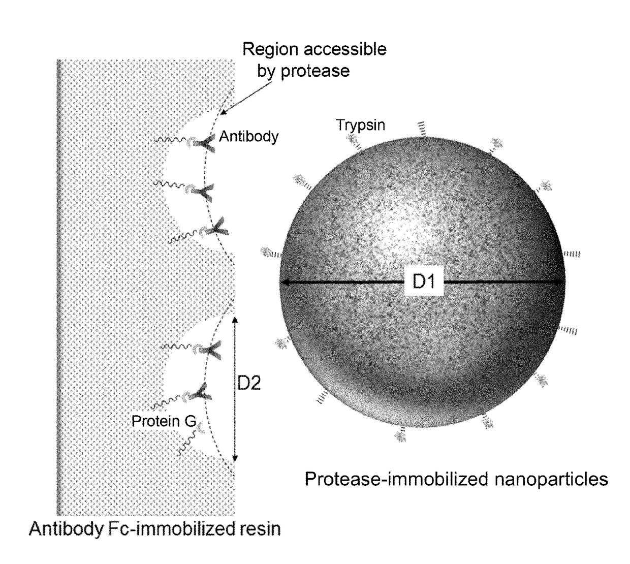
[0190]A kit structured as follows is prepared for analyzing a single sample.[0191](1) PBS buffer (PBS+0.1% n-octyl-β-D-thioglucoside, Dojindo)[0192](2) enzyme reaction buffer (25 mM Tris-HCl, pH 8.0)[0193](3) enzyme reaction termination solution (10% formic acid)[0194](4) filter tube (low-binding hydrophilic PVDF, pore size 0.2 μm, Millipore)[0195](5) low adsorption tube (Richell Microresico® tube 92017)[0196](6) LC-MS vial, insert (Shimadzu GLC, GLC4010-VP, Target vial VP, Target Polyspring insert C4010-630P)[0197](7) porous body (Pierce Protein G UltraLink Resin 53126, 40 μL dispense)[0198](8) nanoparticles (trypsin immobilized FG beads (trypsin 40 μg))[0199](9) instructions

[0200]1. Take 20 μL of blood sample in low adsorption tube (5), and dilute it with 180 μL of buffer (1).[0201]2. Centrifuge porous body (7) using a tabletop-type centrifugal device and discard the supernatant; add 100 μL of buffer (1), gently stir and centrifuge the mixture; discard the supernatant...

example 2

n Filtration Membrane

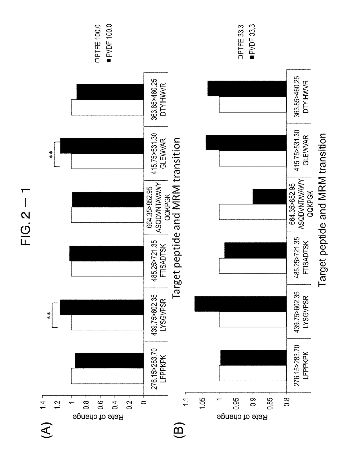
[0215]To study peptide recovery rates when different membrane materials are used for filtration, two membrane materials—polytetrafluoroethylene (PTFE) and polyvinylidene difluoride (PVDF)—were compared.

[0216]The study on peptide recovery rates started with 100 μg / mL trastuzumab, which was then diluted by 3-fold serial dilution (N=4). Protease reaction was performed using nanoparticles with immobilized trypsin (Trypsin TPCK treated). Then, relative to peptides with known sequences (LFPPKPK (SEQ ID NO: 46), LYSGVPSR (SEQ ID NO: 21), FTISADTSK (SEQ ID NO: 3), ASQDVNTAVAWYQQKPGK (SEQ ID NO: 47), GLEWVAR (SEQ ID NO: 6), DTYIHWVR (SEQ ID NO: 5)), recovered peptides were quantified through mass spectrometry (LCMS-8040, Shimadzu Corporation). FIG. 2-1 through FIG. 2-3 show the recovery results of PVDF relative to those of PRFE (rate of variation). In significant difference testing, T-test was employed; (*) was marked for p<0.05 and (**) for p<0.01.

[0217]As a result, no ...

example 3

n Filtration Membrane

[0218]The same test as in Example 2 was conducted using bevacizumab. The results are shown in FIG. 3-1 through FIG. 3-3.

[0219]As a result, the same as in Example 2, no difference in the results was observed in recovery rates among high-concentration peptides, whereas a significant difference was evident in the recovery rates among low-concentration peptides. Accordingly, PVDF was found to be suitable as a filtration membrane.


PUM

PropertyMeasurementUnit
molecular weightaaaaaaaaaa

Abstract

A sample preparation kit related to the present invention provides a significantly versatile analytical technique that is not affected by the diversity of antibodies, difference in species, matrix and the like. For preparing a sample to be used for detection of a monoclonal antibody through high-performance liquid chromatography-mass spectrometry (LC-MS), the kit includes a porous body for immobilizing a monoclonal antibody to be detected; nanoparticles with an immobilized protease; a reaction vessel for selectively digesting the monoclonal antibody by bringing the porous body and nanoparticles into contact; a buffer to be introduced into the reaction vessel along with the nanoparticles and porous body so that a protease reaction is carried out; and a filtration membrane to remove the porous body and nanoparticles after the proteolysis so as to extract the reaction product and the buffer.

Description

TECHNICAL FIELD[0001]The present invention relates to a kit for preparing a sample for detection of a monoclonal antibody through mass spectrometry; more particularly, to such a kit that enables selective protease reaction to obtain peptide fragments containing the specific sequence of a monoclonal antibody so that even more efficient analysis is achieved.BACKGROUND ART[0002]The hardest challenge in drug discovery is to develop drugs capable of showing high efficacy with minor side effects. For that matter, the current focus is on pharmacokinetics, especially therapeutic drug monitoring (TDM). Whether the prescribed dosage is proper and whether the drug has reached the lesion are indicators for drug seeds to be screened so as to determine which drug seeds need to be dropped at an early stage of development. The TDM information is useful in early-stage drug discovery and clinical testing. Personalized medicine, also referred to as order-made medicine, was anticipated at one time; how...

Claims


Application Information

Patent Timeline
Patent Type & Authority Applications(United States)
IPC IPC(8): G01N33/68G01N30/72G01N33/577
CPCG01N33/6848G01N30/7233G01N33/577G01N2333/901C12Q1/00C12Q1/37C12N11/02G01N30/06G01N1/10G01N2030/067G01N2030/8831G01N2030/065G01N27/623
Inventor SHIMADA, TAKASHIIWAMOTO, NORIKO
Owner SHIMADZU CORP
Why Eureka
Try Eureka