
Compositions and methods for delivering an agent to a wound

Inactive Publication Date: 2017-09-14
UNIVERSITY OF MEMPHIS RESEARCH FOUNDATION
View PDF10 Cites 5 Cited by
  • Summary
  • Abstract
  • Description
  • Claims
  • Application Information

AI Technical Summary

Benefits of technology

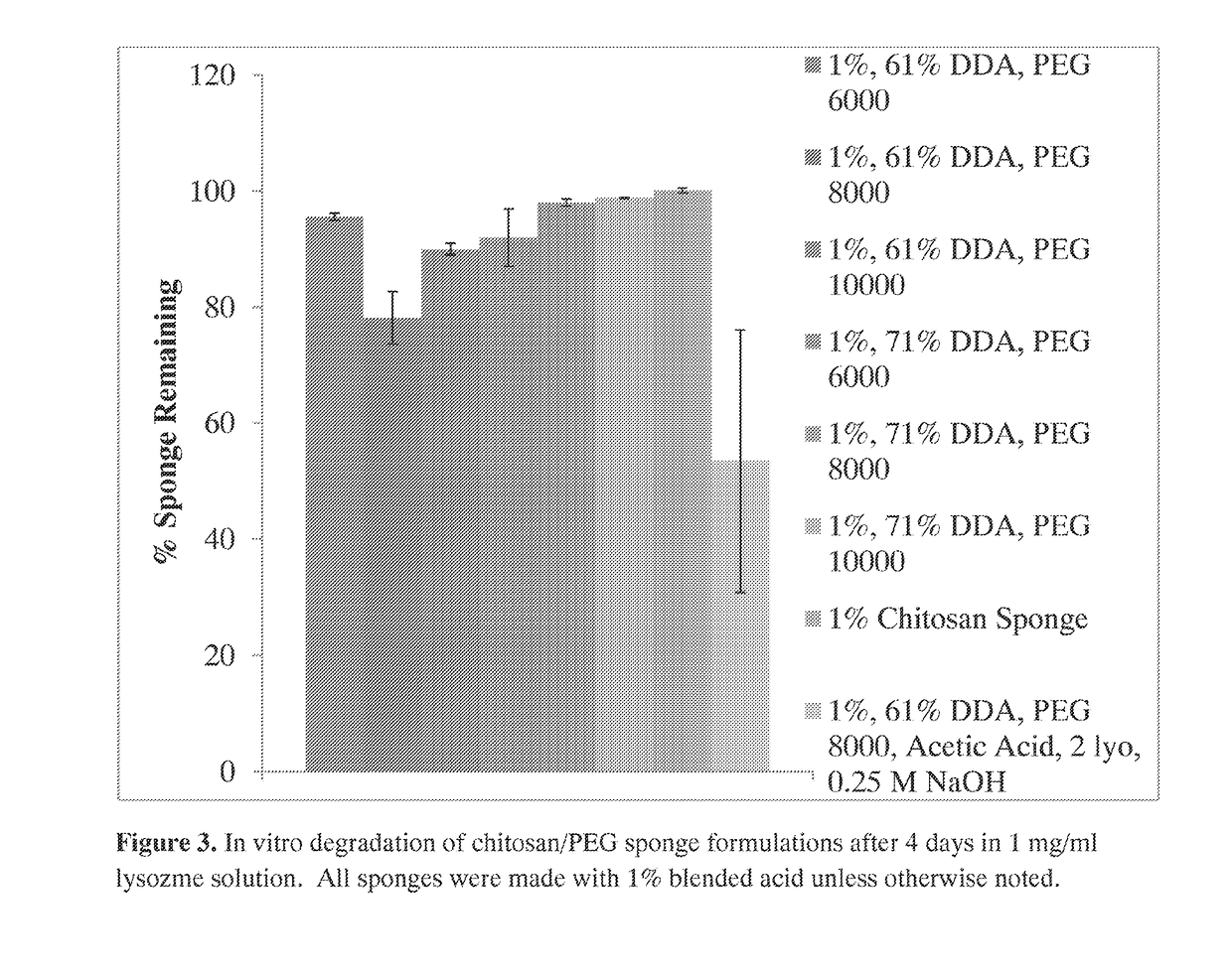
The patent text describes a method to create a chitosan / PEG composition by adjusting the molecular weight of the PEG molecule. By increasing or decreasing the molecular weight, the degradation profile of the composition can be controlled. The text also mentions using analog molecules that have similar functions or structures to the chitosan polymer, which can enhance the function of the composition. The term "prophylactic treatment" refers to reducing the risk of developing a disorder or condition in a person who does not have it.

Problems solved by technology

Musculoskeletal injuries are some of the most prevalent injuries in both civilian (e.g., open fractures) and military (e.g., extremity injuries) populations and their infections can be difficult to treat, often resulting in multiple surgeries and increased costs.
Fungal infections have recently been increasingly problematic in complex extremity trauma and have significantly higher amputation rates than those from bacterial infections.
These problematic, often polymicrobial, infections can result in high healthcare costs, high mortality rates, and significantly higher amputation rates than those from bacterial infections alone.
Wound infections may also be complicated by the presence of multiple bacteria and fungi and by the ability of microbes to form biofilms, which raise the minimum inhibitory concentration (MIC) of antimicrobial agents.
While many local antibiotic delivery systems exist on the market, there are no commercially available local antifungal delivery systems.
However, development of local antifungal delivery systems has been minimal, typically due to the hydrophobicity of antifungals.
These hydrophobic antifungals can be difficult to incorporate into and release from more hydrophilic local drug delivery systems.
Because current methods for treating or preventing infection, particularly fungal infections related to musculoskeletal injuries, are inadequate, compositions and methods for providing agents to prevent or treat an infection at a site of trauma are urgently required.

Method used


Image

  • Compositions and methods for delivering an agent to a wound
  • Compositions and methods for delivering an agent to a wound
  • Compositions and methods for delivering an agent to a wound

Examples


example 1

ic Antifungal Agents are Loaded and Released from Chitosan-PEG Sponges

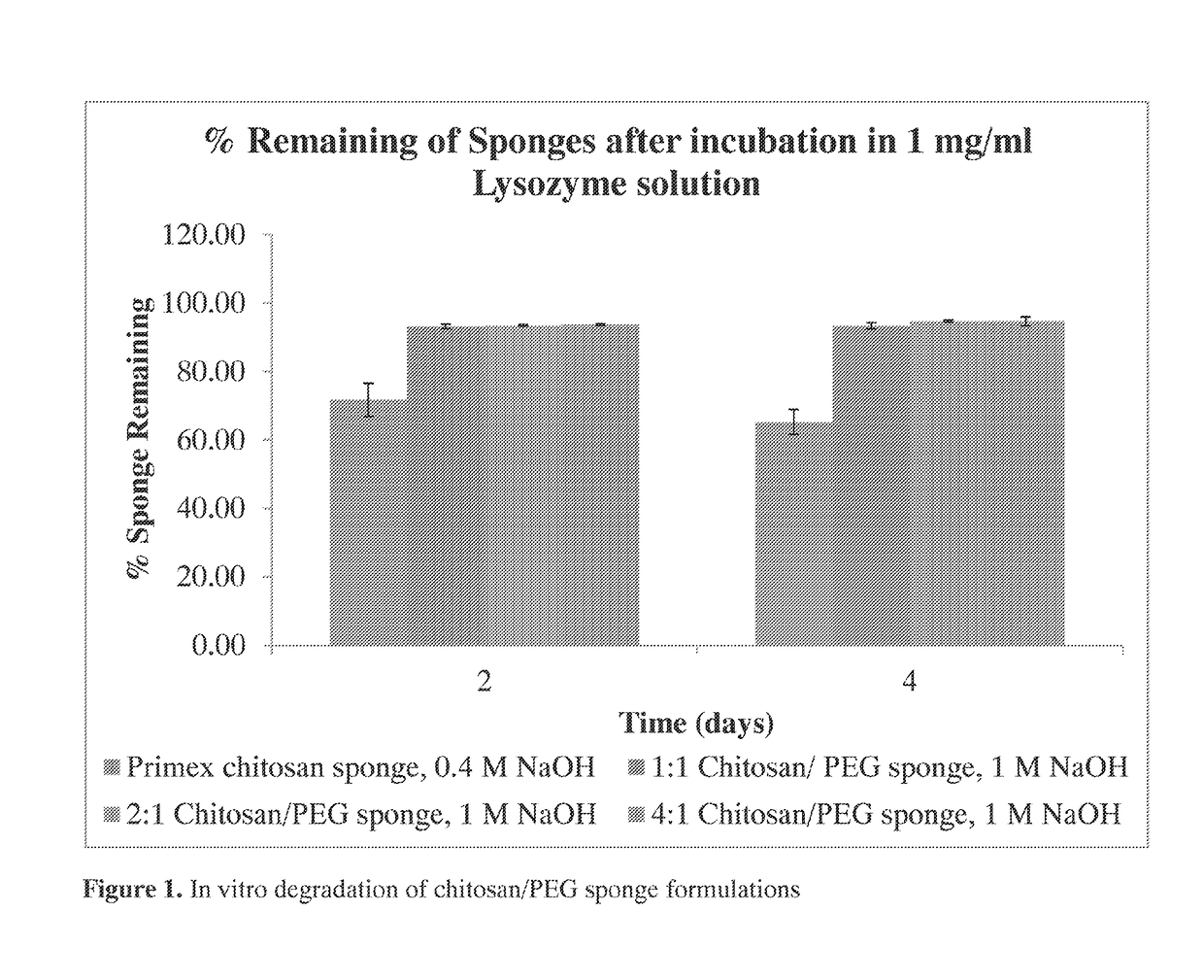
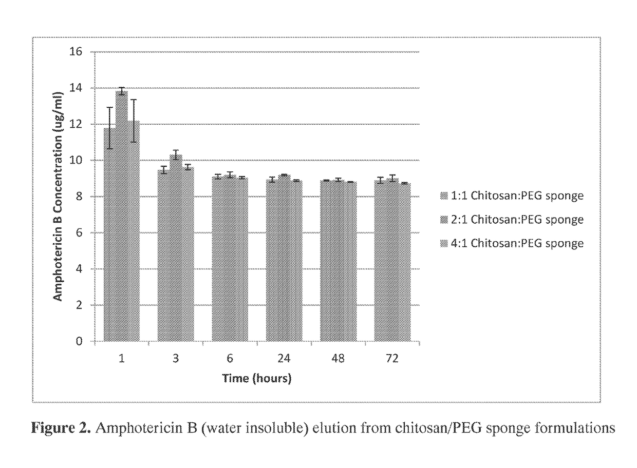
[0180]Polymicrobial musculoskeletal infections, especially from invasive bacteria, fungi or biofilm pathogens, are challenging complications that often increase patient morbidity, mortality, and hospitalization costs (Murray et al., J Trauma 64:S239-251). The clinical need for degradable local antimicrobial delivery systems to fight localized bacteria, fungi, and / or biofilms with patient-tailored antimicrobial point-of-care loading has resulted in the previous development of a chitosan sponge local delivery system (Hanssen, Clin Orthop Relat Res:91-96; Noel et al., Clin Orthop Relat Res 468:2074-2080; Stinner et al., J Orthop Trauma 24:592-597). The present study was designed to determine if the blending of these chitosan sponges with polyethylene glycol would result in local drug delivery systems capable of antifungal and antibiotic release for the prevention of polymicrobial wound infections.

[0181]A set of chito...

example 2

rier Transform Infrared Spectroscopy) Analysis of Chitosan / PEG Blended Sponges

[0235]FTIR (Fourier transform infrared spectroscopy) analysis of the chitosan / PEG blended sponges and control chitosan sponges was performed (FIG. 11). The presence of PEG in the sponges was confirmed by FTIR. Characteristic peaks were seen in the 1% chitosan sponges at 3290 cm−1 (O—H stretching), 1643 cm−1 (amide I C═O stretching), and 1557 cm−1(amide II N—H) (Wang et al. J Biomed Mater Res Part A 2008; 85(4):881-7; Kolhe et al. Biomacromolecules 2003; 4(1):173-180; He et al., Chin J Polym Sci 2009; 27(4):501-510). Characteristic C—O bending peaks were observed in PEG 6000 and 8000 at 1093 cm−1, and peaks from the crystalline region of PEG were observed at 1278, 960, and 840 cm−1 (Wang et al. J Biomed Mater Res Part A 2008; 85(4):881-7; Kolhe et al. Biomacromolecules 2003; 4(1):173-180).

[0236]All blended sponges exhibited the characteristic chitosan O—H stretching peaks at 3357-3360 cm−1. All peaks were s...

example 3

fraction (XRD) Analysis of Chitosan / PEG Blended Sponges

[0238]X-ray diffraction results revealed a difference between the chitosan powder and all sponge types (FIG. 12A). Analysis of the XRD spectra of the chitosan powder indicated the presence of anhydrous chitosan crystal structure (Ogawa et al., Agric Biol Chem 1991; 55(9):2375-2379). The chitosan powder exhibited the typical crystalline peak at approximately 20°, but this peak disappeared upon manufacturing the powder into sponges, both in the control chitosan sponges or the chitosan / PEG blended sponges. After chitosan dissolution and lyophilization, the crystalline peak at 20° disappeared and the 10° peak decreased and became broader, suggesting a large loss in helical crystalline forms (Zhang et al., Carbohydr Res 2005; 340(11):1914-7). Additionally, the peak at approximately 12° in the chitosan powder decreased upon sponge manufacturing, and the 1% chitosan, chitosan / PEG 6000 2 lyo, and chitosan / PEG 8000 1 lyo sponges displaye...


PUM

PropertyMeasurementUnit
Fractionaaaaaaaaaa
Timeaaaaaaaaaa
Timeaaaaaaaaaa

Abstract

The invention provides compositions featuring chitosan and polyethylene glycol and methods for using such compositions for the local delivery of biologically active agents to an open fracture, complex wound or other site of infection. Advantageously, the chitosan-PEG compositions can be loaded with one or more antimicrobial agents, including hydrophobic agents, and can be tailored to the needs of particular patients at the point of care (e.g., in a surgical suite, clinic, physician's office, or other clinical setting).

Description

CROSS REFERENCE TO RELATED APPLICATIONS[0001]This application claims benefit of U.S. Provisional Application Ser. No. 61 / 940,296, filed Feb. 14, 2014, and 62 / 077,047, filed Nov. 7, 2014, the contents of each of which are incorporated herein by reference in their entirety.STATEMENT OF RIGHTS TO INVENTIONS MADE UNDER FEDERALLY SPONSORED RESEARCH[0002]This invention was made with U.S. government support under grant no. DM090455 awarded by United States Army Medical Research Acquisition Activity (USAMRRAA); Grant Nos.: W81CWH-09-PROP-HAD and W81XWH-12-2-0020 awarded by United States Army Medical Research and Materiel Command (USAMRMC); Grant Nos. W81XWH-08-1-0312, Proposal No. 07128022, and W81XWH-12-2-0020, Proposal No. DM090455 awarded by the Department of Defense; and Grant No. 2009087439 awarded by the National Science Foundation. The U.S. government has certain rights in the invention.BACKGROUND OF THE INVENTION[0003]Musculoskeletal injuries are some of the most prevalent injuries ...

Claims


Application Information

Patent Timeline
IPC IPC(8): A61L29/16A61K47/10A61L29/08A61L29/04A61L29/14C08J5/18A61K9/00A61K38/14A61K31/7036C08L5/08C08L71/02A61K47/36A61K31/7048
CPCA61L29/16C08J2371/02A61K47/10A61L29/085A61L29/049A61L29/146A61K31/7048A61K9/0024A61L29/148A61K38/14A61K31/7036C08L5/08C08L71/02C08J5/18A61L2300/404A61L2300/232A61L2300/252A61L2420/06A61L2300/606C08L2203/02C08J2305/08A61K47/36C08J9/0061C08J9/28C08J2201/0484C08J2205/022C08J2207/10C08J2471/02A61L15/26A61L15/28A61L15/425A61L15/44A61L17/005A61L17/06A61L17/10A61K9/19
Inventor HAGGARD, WARREN O.PARKER, ASHLEY C.JENNINGS, JESSICA AMBER
Owner UNIVERSITY OF MEMPHIS RESEARCH FOUNDATION
Learn More