
Porous Polymer Scaffold Useful for Tissue Engineering in Stem Cell Transplantation

a porous polymer and stem cell technology, applied in the field of biodegradable porous polymer scaffolds, can solve the problems of limited tissue availability, morbidity and mortality, artificial skin embedded with epidermal/fibroblastic cells, etc., and achieve the effect of increasing the engraftment of male bmscs

Active Publication Date: 2017-04-27
COUNCIL OF SCI & IND RES
View PDF1 Cites 1 Cited by
  • Summary
  • Abstract
  • Description
  • Claims
  • Application Information

AI Technical Summary

Benefits of technology

The present invention is about a biodegradable, stable, and biocompatible porous polymer scaffold that can be used to deliver cells to damaged tissue. The scaffold is made from a special type of polymer called polyethyleneglycol-polyurethane (PEG-PU) and castor oil cross linkages. The scaffold is evaluated in various cell lines and primary bone marrow stem cells to determine its ability to induce porosity and protect against oxidative stress. The scaffold is also tested in a mouse model to determine its suitability for delivering cells to damaged tissue. The technical effects of this invention include providing a safe and effective scaffold for cell delivery and promoting tissue regeneration.

Problems solved by technology

Chronic wound remains as a constant challenge for patients with diabetes or treated with immunosuppressive drugs, chemo- and radio-therapies.
A complex cascade of regulated steps with complications of persistent inflammation, fibroblast dysfunction and impaired angiogenesis often leads to morbidity and mortality.
807-818), conventional surgical treatments like skin autograft and allograft, artificial skin embedded with epidermal / fibroblastic cells has their own disadvantages such as limited tissue availability, high cost, immune-rejection and prolonged time of healing.
Nevertheless, this strategy, has also met with limited success owing to enhanced ROS (Reactive oxygen species) levels at injury site that impairs the function of stem cells transplanted at chronic wounds (see, Case, J. et al Antioxid.
However, contradictory reports exist regarding the biocompatible properties of these PLA-PGA-based biopolymers, (see, Makadia, H. K et al., Polymers 2011, 3, pp.
However, the mechanisms of cell penetration into these polyurethane scaffolds are lacking in literature.
While biodegradation in polyurethanes can be easily achieved by incorporating hydrolysable moieties like esters in the polymeric chain, control on occasional cytotoxicity of cleaved fragments post-degradation remains the key area of concern and challenge.
Often small molecules with three or more functional groups (f>3) conventionally used as cross-linkers can lead to increased toxicity upon degradation.
One way to troubleshoot is using larger molecules with active functional groups, which however in most cases can introduce a lot of hard segments difficult to be fragmented under mild conditions.

Method used


Image

  • Porous Polymer Scaffold Useful for Tissue Engineering in Stem Cell Transplantation
  • Porous Polymer Scaffold Useful for Tissue Engineering in Stem Cell Transplantation
  • Porous Polymer Scaffold Useful for Tissue Engineering in Stem Cell Transplantation

Examples


example 1

of Polymer Networks

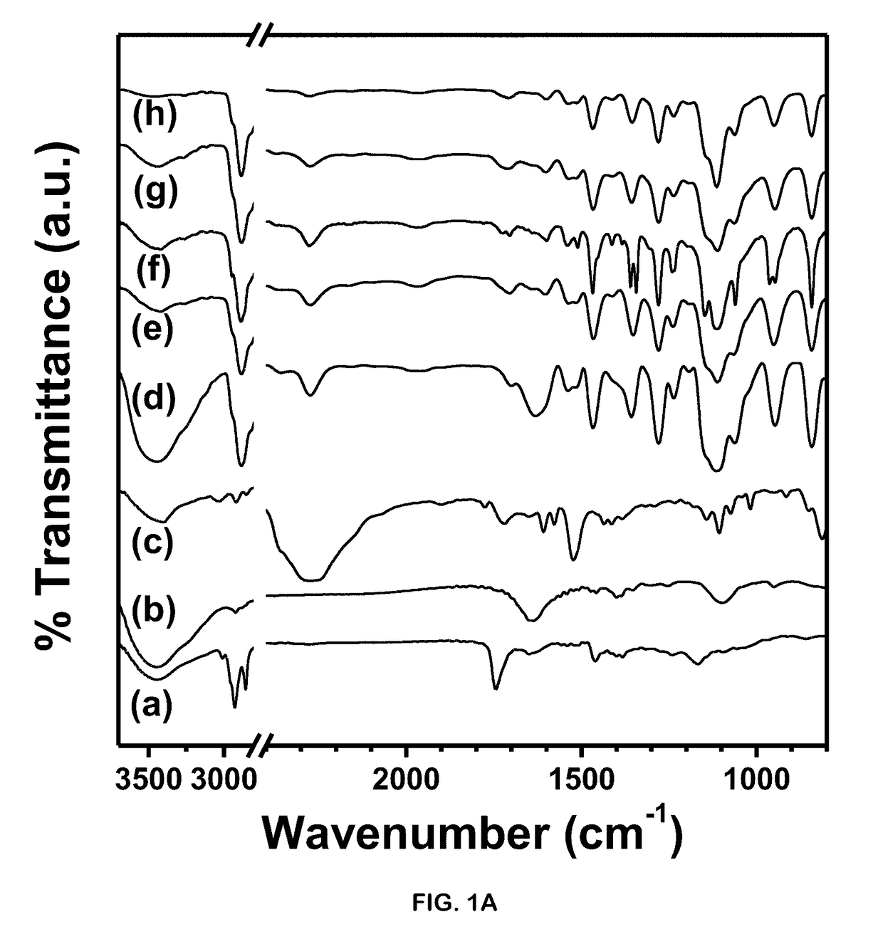
[0103]Polyethyleneglycol-polyurethane networks when subjected to autoclaving (121° C. temperature and 15 psi pressure) did not exhibit any significant changes confirming high thermo- and barostablity (FIG. 1E). Often suitability of the polymer network depends upon its biodegradability and rate of release of implanted cells to interact with in vivo microenvironment. In pathological conditions, cellular damage including injury to lysosomal membranes results in leakage of their enzymes into the cytoplasm and activation of acid hydrolases that leads to acidic intracellular pH. Thus targeting acidic microenvironment of injured tissue often necessitates the delivery vehicles with pH-sensitive degradation in order to release cells at a controlled rate. (See, Stubbs, M et al., Mol. Med. 2000, 6, pp. 9-15). Interestingly, when the networks were subjected to buffers of different pH, they showed an increased degradation at acidic pH 5.8 (2.5-fold percent change in weight) as...

example 2

ability of Polymer Networks

[0104]Polymer degradation in presence of biocatalysts indicates the feasibility of use as cell delivery vehicles in transplantation therapies. Bio-catalytic degradation studies performed for 24 h, revealed 50% degradation in presence of collagen at 1 mg / mL and a relatively less degradation (13%) in presence of enzyme 0.25% trypsin (FIG. 2B). Polymer networks also showed 22.3% degradation over period of 24 h in presence of 20% TCA suggesting chemical hydrolysis by the reducing agent. The results imply that these polymer networks can efficaciously release cells making them a probable cell delivery vehicle.

example 3

ibility of Polymer Networks

[0105]The polymer networks when tested for their cytotoxic effect on cells such as MDA-MB-231, SK-HEP1 and mouse BMSCs depicted an insignificant difference on viability in presence or absence of polymer networks. Trypan blue dye exclusion assay performed to evaluate the stained dead cell population also revealed no significant difference between cells cultured with or without polymer networks (FIG. 2C). Furthermore, SRB Assay performed to evaluate the cytotoxicity imparted to the cultured cells showed an insignificant difference between results of cells cultured in absence or presence of polymer networks (FIG. 2D).


PUM

PropertyMeasurementUnit
Temperatureaaaaaaaaaa
Fractionaaaaaaaaaa
Timeaaaaaaaaaa

Abstract

The present invention relates to the synthesis of porous polymer scaffold from polyethyleneglycol-polyurethane having castor oil linkages under controlled conditions and their use as stem cell delivery vehicles thereby accelerating the tissue regeneration process. The present invention further studies the biodegradability, stability and biocompatibility of porous polymer scaffolds in varios cell lines and primary bone marrow stem cells. Particularly the present invention further relates to the physio-chemical characterization of the porous polymer scaffolds.

Description

PRIORITY CLAIM[0001]The present application claims priority to Indian Application No. 3470 / DEL / 2015 filed Oct. 27, 2015, which is incorporated herein by reference.FIELD OF THE INVENTION[0002]The invention relates to the synthesis of biodegradable porous polymer scaffold from polyethyleneglycol-polyurethane (PEG-PU) under controlled conditions and its use as cell delivery vehicles of stem cells thereby accelerating the tissue regeneration processes, in particular to chronic wounds, diseases related to impaired neo-vascularization and ROS-induced degenerative diseases.BACKGROUND OF THE INVENTION[0003]Chronic wound remains as a constant challenge for patients with diabetes or treated with immunosuppressive drugs, chemo- and radio-therapies. Mechanisms underlying poor wound healing are still unclear (see, Fang, et al., Nutrition. 2002, 18, pp. 872-879). Wound healing is generally characterized by haemostasis, inflammation, migration and proliferation of fibroblasts, endothelial and epit...

Claims


Application Information

Patent Timeline
IPC IPC(8): A61L27/18A61L27/56A61L27/54C08L75/08C08G18/48C08G18/08C08J9/26C08L71/02C12N5/00C08G18/10
CPCA61L27/18C08J2371/02A61L27/56A61L27/54C08G18/10C08G18/4825C08G18/14C08J9/26C08L71/02C08L75/08C12N2533/30C12N2537/10C08J2375/08C08J2201/0462C08J2207/10C08L2205/04C08L2203/02C12N5/0068A61L27/58C08G18/4833C08G18/7671C08G18/8051C08G65/33351C08G18/36C08J2205/044C08J2205/05C08J2300/16
Inventor DAS, AMITAVABASAK, PRATYAYGEESALA, RAMASATYAVENIBAR, NIMAIDHOKE, NEHA RAGHUVIRKAUSHIK, KOMAL
Owner COUNCIL OF SCI & IND RES
Learn More