
Patient Eye Protection Device For Dermatology, X-Ray, and General Anesthesia Procedures

a technology for dermatology, x-ray and general anesthesia, applied in the field of eye protection covers, can solve the problems of presenting potential hazards to the eyes of patients, and affecting the patient's vision, and causing damage to the patient's eyes

Inactive Publication Date: 2017-04-27
SOJO MEDICAL
View PDF0 Cites 0 Cited by
  • Summary
  • Abstract
  • Description
  • Claims
  • Application Information

AI Technical Summary

Benefits of technology

The present invention relates to an eye protection cover for use during medical or health care procedures that may pose a hazard to the eyes of a patient. The cover is made of a heat-resistant material that conforms to the shape of the eye and includes a handle for easy application and removal. The cover is designed to protect the eye from optical energy, such as laser treatment, and also from mechanical or chemical damage. The invention addresses the need for an improved and simple eye protection device that can be used during X-ray procedures and general anesthesia to prevent eye injuries and protect the cornea from abrasions.

Problems solved by technology

Common dermatological and aesthetic procedures such as laser treatments, intense pulsed light, LED therapy, microdermabrasion, and others present potential hazards to the eyes of the patient.
The optical energy from light based devices such as laser, intense pulse light (IPL), and LED therapy present an eye hazard related to the energy inherent in these treatment modalities.
Optical energy from these devices is absorbed by the cornea and / or retina and may damage the vision of the patient.
Other dermatological therapies such as microdermabrasion and chemical treatments can cause mechanical or chemical damage to the eyes of the patient.
Protection of the patient's eyes during dermatological and aesthetic procedures can be challenging, particularly when the procedures are performed around the eyes and face.
Traditional protective eyewear and masks often sit too far away from the eye, leaving a risk of damage to the eyes.
Traditional eyewear is often too big and bulky, and interferes with the treatment.
The Arnold et al. device, however, suffers from a number of shortcomings, and in particular relating to ease of use and effectiveness.
If a laser were to contact this material, it is likely that the energy from the laser would melt the material, and likely burn the patients.
Furthermore, the eye-cover is two dimensional and therefore does not properly confirm to the contour of the eyes, which causes coverage issues and issues relating to comfort.
The adhesive on this device sticks to the gloves (or to a person's hands even if gloves were not worn) making the device difficult to apply and difficult to reposition on the patient's eyes.
As mentioned, the device is planer in nature and fails to adequately follow the three dimensional contours of the eye.
Also, the device is difficult to handle since it requires touching the adhesive, which sticks especially to gloves.
It is known that exposure to ionizing radiation can lead to cataracts.
X-ray radiation, especially repeated exposure, can lead to other unwanted complications.
While a number of more serious eye injuries can result from general anesthesia, the most common injury is corneal abrasion.
Corneal abrasions can cause mild to severe discomfort, irritation, and pain.
Additionally, corneal abrasion may also effect normal vision.
The dry cornea may stick to the inside of the eyelid, causing an abrasion when the eye opens post surgery.
Corneal abrasion may also occur if something brushes against the open eye during surgery.
These measures and devices, however, are not always sufficient, because corneal abrasions and other eye injuries associated with surgery and medical procedures are still common.
Furthermore, the application of gauze and tape to protect the eye makes it impossible for the anesthesiologist to easily view the patient's pupils, which is an important part of anesthesia management.
Despite these best efforts injuries and irritation do take place.
Simply using gel and gauze pads still fails to eliminate post surgical eye redness, irritation and pain in up to 50% of surgical cases.
These symptoms, typically resolve in a matter of hours, but can be range from mildly to very irritating and painful.

Method used


Image

  • Patient Eye Protection Device For Dermatology, X-Ray, and General Anesthesia Procedures
  • Patient Eye Protection Device For Dermatology, X-Ray, and General Anesthesia Procedures
  • Patient Eye Protection Device For Dermatology, X-Ray, and General Anesthesia Procedures

Examples


Embodiment Construction

[0028]The purpose of the present invention, in its various forms, is to protect the eyes of a patient undergoing dermatological procedures, x-ray procedures, general anesthesia, as well as other treatments such as those common to health spas such as tanning, facials, and other similar and related fields of use.

[0029]The invention consists of an injection molded silicone eye cup, or cover. The eye cover is three dimensionally shaped to fit within the eye socket of the patient.

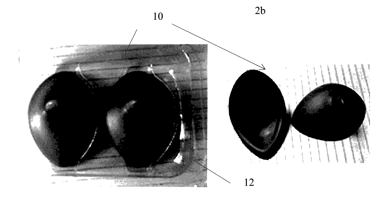
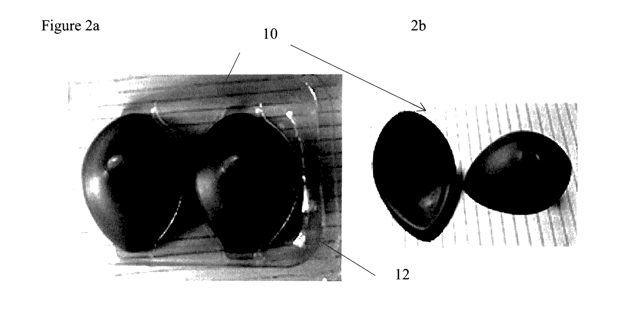
[0030]FIG. 2a shows a top view of the eye cover 10. The cover is initially provided releaseably secured to a casing 12, such as the plastic casing shown in FIG. 2a. The eye cover can be removed from the casing just prior to application. Alternatively, they eye cover 10 can be packaged in a heat sealed foil pouch, which eliminates the need for a releasably secured casing. The outer portion of the inside edge of the eye cover 10 is coated with an adhesive, which will also hold the eye cover 10 in place when applie...


PUM


Abstract

The present invention relates to an eye protection cover; and in particular, to an eye protection cover that protects the eye during medical or health care treatment procedures. The cover is comprised of a heat resistant material, conforms in three dimensions to the shape of the eye, and includes a handle for ease of insertion and removal.

Description

RELATED APPLICATIONS[0001]The present application claims priority to and incorporates by reference U.S. Provisional Patent Application No. 62 / 019,650 filed on Jul. 1, 2014. The present application incorporates by reference U.S. patent application Ser. No. 13 / 786,897 filed on Mar. 6, 2013.BACKGROUND OF THE INVENTION[0002]Field[0003]The present invention relates to an eye protection cover; and in particular, to an eye protection cover that protects the eye during medical or health care treatment procedures. The cover is comprised of a heat resistant material, conforms in three dimensions to the shape of the eye, and includes a handle for ease of application or insertion and removal.[0004]Background[0005]Dermatology & Aesthetics[0006]Common dermatological and aesthetic procedures such as laser treatments, intense pulsed light, LED therapy, microdermabrasion, and others present potential hazards to the eyes of the patient.[0007]The optical energy from light based devices such as laser, ...

Claims


Application Information

Patent Timeline
Patent Type & Authority Applications(United States)
IPC IPC(8): A61F9/04B65D73/00
CPCB65D73/0028A61F9/045A61F9/04
Inventor O'BRIEN, JOE
Owner SOJO MEDICAL
Learn More