High resolution imaging mass spectrometry

Active Publication Date: 2017-03-23
BAYLOR UNIVERSITY
View PDF6 Cites 0 Cited by
  • Summary
  • Abstract
  • Description
  • Claims
  • Application Information

AI Technical Summary

Benefits of technology

This patent describes a system and method for mass spectrometry imaging that uses different techniques for desorption and ionization. The system can analyze samples directly without the need for a matrix and has the resolution to analyze molecules in nano-meters. The method allows for softer desorption using a larger ionization cross-section in the second stage, resulting in molecular and fragment ions for analysis. Additionally, the method has increased spatial resolving power, compatibility with various mass analyzers and detectors, and allows for increased sensitivity and resolution with small spot sizes.

Problems solved by technology

A known issue with MALDI is that the application of the matrix substance to the sample tissue can cause migration of small cells and / or sample components that affect the accuracy of an analysis on a cell-by-cell resolution.
Moreover, MALDI matrix may interact with surface chemicals and lead to bias analysis of certain classes of compounds.
Further, a MALDI technique is generally only capable of probing in the 5-50 micro-meter (μm) range due to the large dimensions of the laser spot caused by a light diffraction limit,
SIMS can be too destructive to the sample to analyze various aspects of the sample.
Mile advances have been made in IMS, there are currently no known methods for desorbing intact molecules from smaller than one micron surfaces using soft ionization approaches.
Although MALDI can desorb and concurrently ionize with a laser and an applied matrix for soft desorption, the resolution of a MALDI process is too large for cell-by-cell or particularly subcellular structures or other small samples that may be dislodged or otherwise desorbed for analysis by a mass spectrometer.
SIMS has the capability of such small resolution, but with its hard ionization, can result in fragmentation that disrupts the structure of the molecule that is being sought to analyze.
However, radio frequency ionization (“RFI”) may not have the resolution to directly desorb smaller selected portions of a sample.

Method used


Image

  • High resolution imaging mass spectrometry
  • High resolution imaging mass spectrometry

Examples


Embodiment Construction

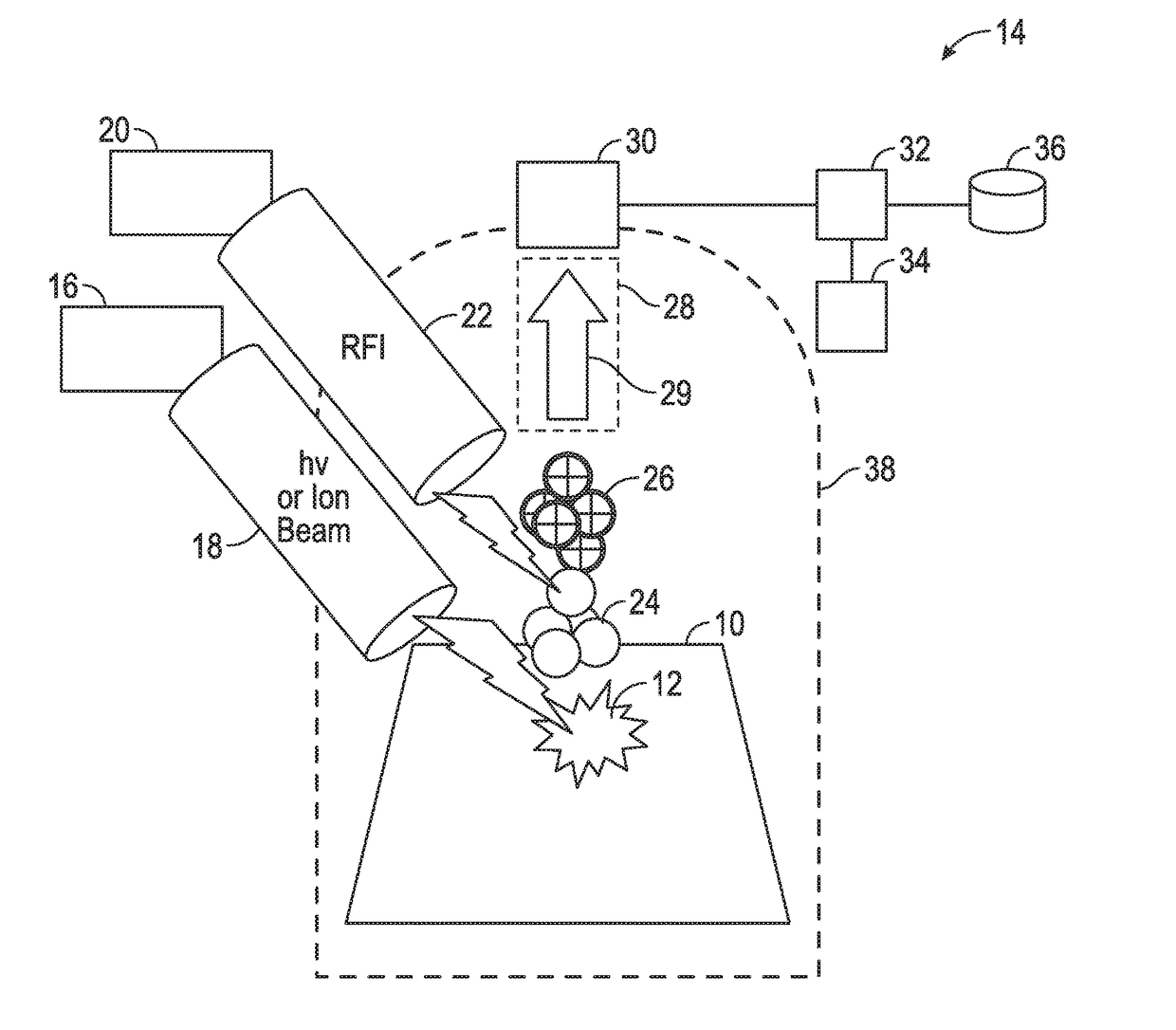
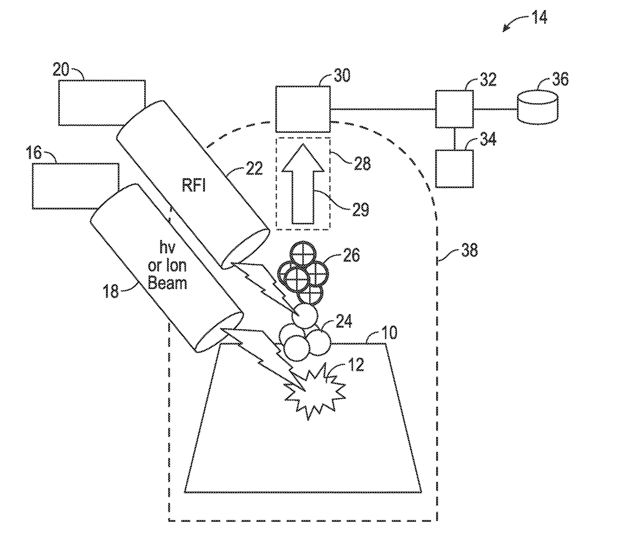
[0021]The FIGURE described above and the written description of specific structures and functions below is not presented to limit the scope of what Applicant has invented or the scope of the appended claims. Rather, the FIGURE and written description are provided to teach any person skilled in the art to make and use the inventions for which patent protection is sought. Those skilled in the art will appreciate that not all features of a commercial embodiment of the is inventions are described or shown for the sake of clarity and understanding, Persons of skill in this art will also appreciate that the development of an actual commercial embodiment incorporating aspects of the present disclosure will require numerous implementation-specific decisions to achieve the developer's ultimate goal for the commercial embodiment, Such implementation-specific decisions may include, and likely are not limited to, compliance with system-related, business-related, government-related and other con...


PUM


Abstract

The present disclosure provides a system and method for mass spectrometry imaging in a multi-stage ionization applying different technologies by decoupling the desorption and ionization events. At a first stage, a primary beam, such as an ion beam, desorbs one or more molecules of a targeted sample, and at a second stage the desorbed molecules are ionized. The system and method can act independent of a matrix application to the target sample for a direct analysis and has the spatial resolution needed to operate in nano-meters resolution for a cell-by-cell analysis, if desired. The first stage desorption applies a first technique that allows neutral molecules of the target sample to become desorbed from the surface without requiring the molecules to be ionized during the desorption. The second stage ionizes the neutral molecules after the desorption in the first stage, when the defined target molecules have been volatilized.

Description

CROSS REFERENCE TO RELATED APPLICATIONS[0001]This application claims the benefit of U.S. Provisional Application No. 62 / 220,452, filed Sep. 18, 2015, which is incorporated by reference herein.STATEMENT REGARDING FEDERALLY SPONSORED RESEARCH OR DEVELOPMENT[0002]Not applicable.REFERENCE TO APPENDIX[0003]Not applicable.BACKGROUND OF THE INVENTION[0004]Field of the Invention[0005]The disclosure generally relates to mass spectrometry. More specifically, the disclosure relates to high resolution imaging mass spectrometry.[0006]Description of the Related Art[0007]Mass spectrometry (“MS”) is increasingly being used to analyze the composition of materials. Literature generally describes MS as an analytical technique that produces spectra of the masses of the atoms or molecules of a sample of material. The spectra are used to determine the elemental compositions or isotopic signatures of a sample, the masses of particles and of molecules, and if applicable, the chemical structures of molecule...

Claims


Application Information

Patent Timeline
IPC IPC(8): H01J49/00H01J49/10
CPCH01J49/0004H01J49/0031H01J49/105H01J49/0463
Inventor SOLOUKI, TOURADJ
Owner BAYLOR UNIVERSITY
Why Eureka
Try Eureka