Activators of myosin ii for modulating cell mechanics
a technology of activated myosin and cell mechanics, applied in the field of compounds as activated myosin ii, can solve the problems of each being impaired by molecules that modulate the cell's mechanical machinery, and achieve the effects of increasing cell tension and elasticity, and promoting myosin ii accumulation
- Summary
- Abstract
- Description
- Claims
- Application Information
AI Technical Summary
Benefits of technology
Problems solved by technology
Method used
Image
Examples
example 1
CIMPAQ Processes of High-Throughput Data and Identification of Mechanical Modulators, Mitotic Inhibitors, and Lethal Compounds
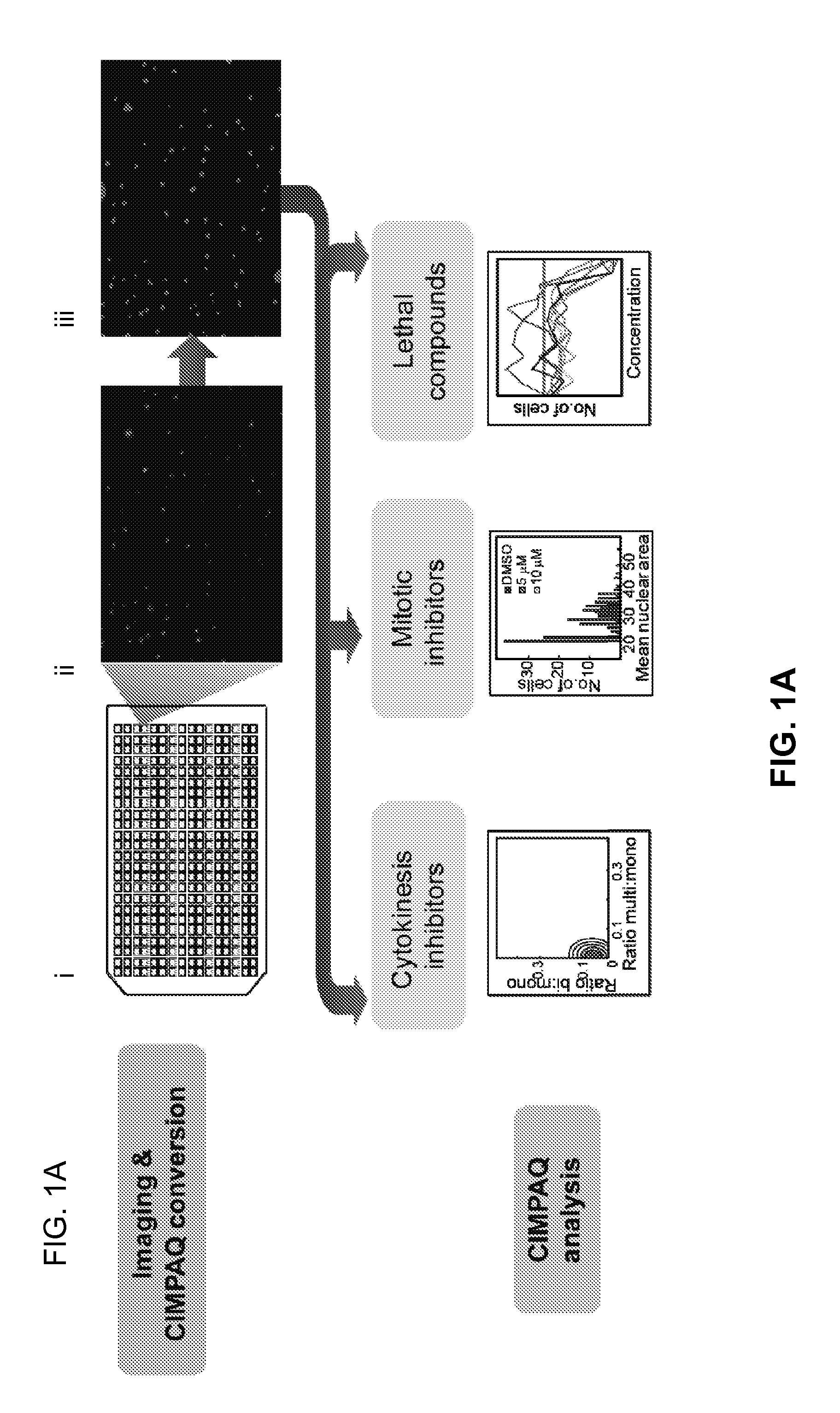
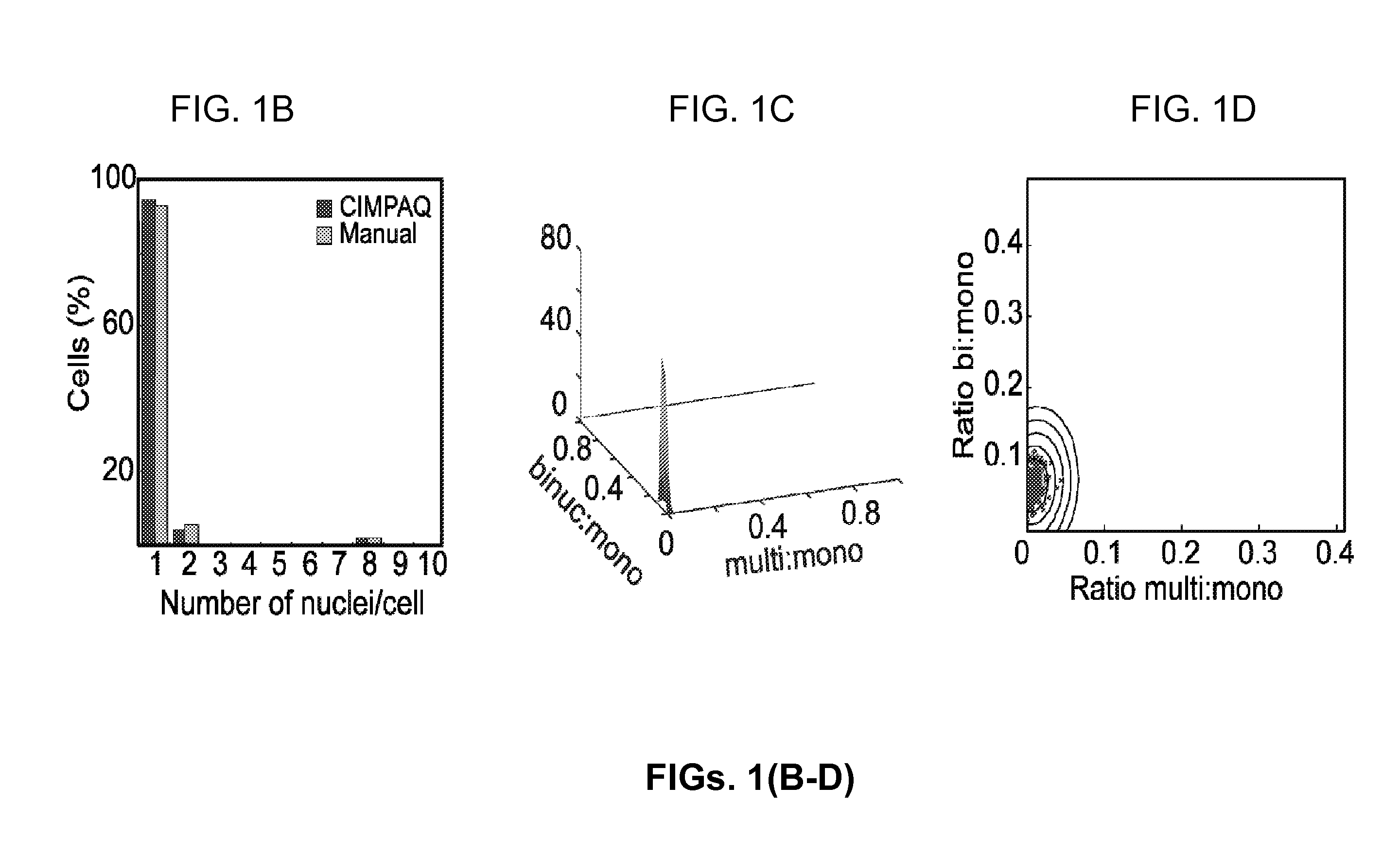
[0110]FIGS. 1(A-D) are a set of diagrams and graphs showing CIMPAQ processes of high-throughput data and identification of mechanical modulators, mitotic inhibitors, and lethal compounds. FIG. 1A shows workflow diagram of primary screening from 384-well plating (i) to raw data acquisition (ii) to CIMPAQ image conversion by segmentation (iii). Cytokinesis hits are identified in a 5-step process: Acquisition of FIG. 1A(ii) raw images of NLS-tdTomato expressing cells and conversion into FIG. 1A (iii) CIMPAQ-processed version FIG. 1B shows sample histogram of a single well showing the distribution of nuclei per cell counts demonstrating high agreement between manual counts and CIMPAQ analysis. The Cartesian coordinates defined by the ratio of bi- to mono-nucleated cells and the ratio of multi- to mononucleated cells of the untreated WT wells are fitted to a two d...
example 2
Identification of Carbamate-7 as a Cytokinesis Inhibitor Affecting the Myosin II-RacE Pathway
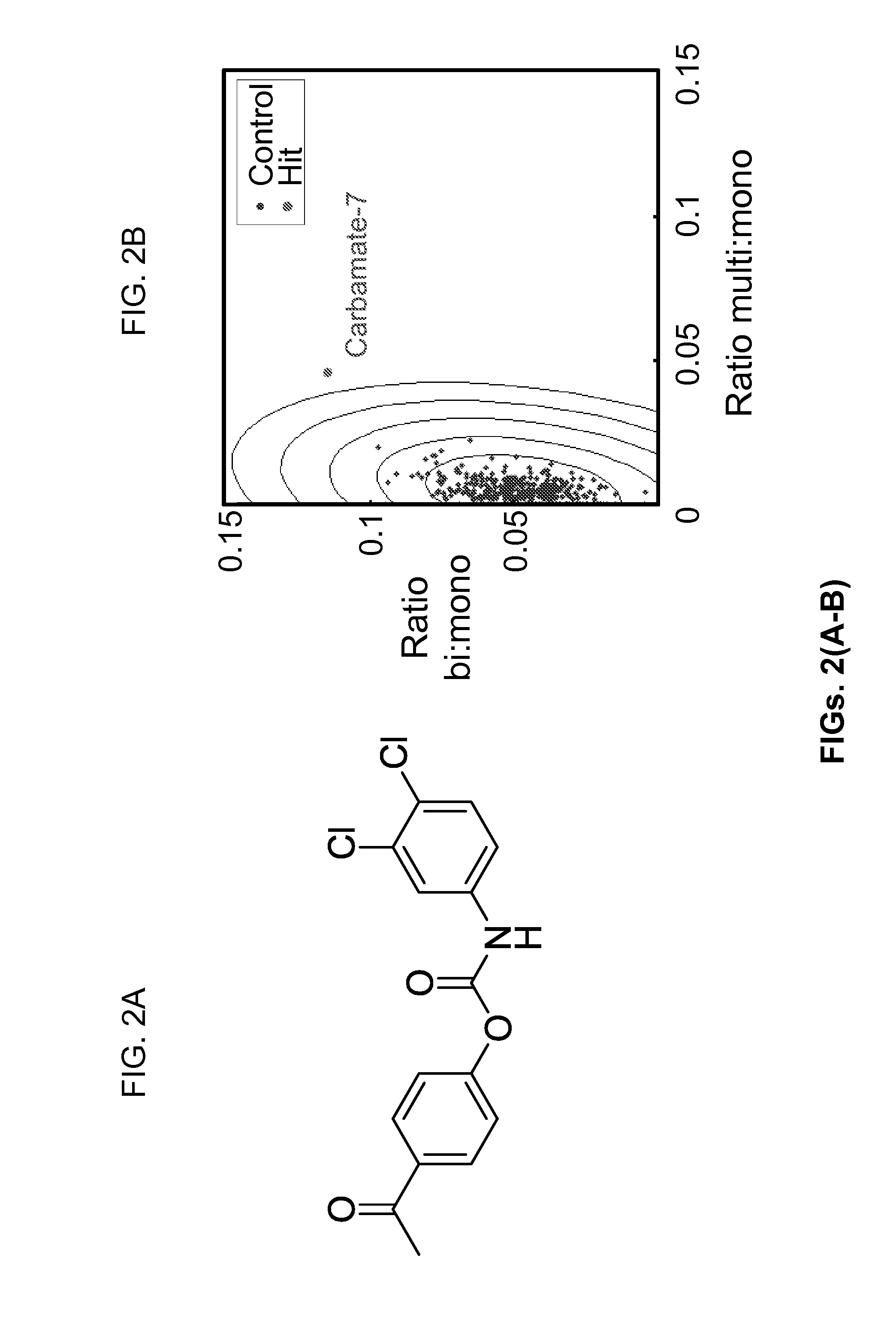
[0111]FIGS. 2(A-D) are a set of images and graphs showing the molecular structure of carbamate-7 and identification of carbamate-7 as a cytokinesis inhibitor affecting the myosin II-RacE pathway according to one embodiment of the present invention. FIG. 2A shows the structure of the putative carbamate-7. In FIG. 2B, cells treated with carbamate-7 (red) showed a shift in the nuclei / cell distribution over six standard deviations from the control data (blue), in primary screening. FIG. 2C shows that partial dose response curves reveal that carbamate-7 increases the fraction of binucleates at nM concentrations. In FIG. 2D, results from synthetic lethality experiments show a statistically significant difference in the average number of nuclei / cell between untreated and treated samples in wild-type and kif12 null strains (**p<0.0001), but not myoII or RacE null strains. Error bars represent SEM.
example 3
Myosin II Cortical Dynamics Affected by Treatment with Carbamate-7
[0112]FIGS. 3(A-D) are a set of images and graphs showing that myosin II cortical dynamics affected by treatment with carbamate-7 according to one embodiment of the present invention. FIG. 3A: Structural Illuminated Micrographs of myoII:GFP myoII cells show an increase in the amount and variability of myosin II bipolar thick filaments in 500-nM carbamate-7 treated (right panels) versus untreated (left panels) cells. In both, the white box represents a zoomed in region, shown to the right of the main images. FIG. 3B: Total Internal Reflection Microscopy (TIRF) images of cells treated with increasing amounts of carbamate-7 show increase of cortical GFP-myosin II, quantified in FIG. 3C. FIG. 3D: Sedimentation assay shows increase of non-monomeric myosin II in 700-nM carbamate-7 treated over untreated cells (n=3). FIG. 3E: Cortical tension measurements show a 1.4-fold increase in cells acutely treated with carbamate-7. Er...
PUM
| Property | Measurement | Unit |
|---|---|---|
| temperature | aaaaa | aaaaa |
| mechanical | aaaaa | aaaaa |
| catalytic activity | aaaaa | aaaaa |
Abstract
Description
Claims
Application Information
 - R&D
- Intellectual Property
- Life Sciences
- Materials
- Tech Scout
- Unparalleled Data Quality
- Higher Quality Content
- 60% Fewer Hallucinations
Browse by: Latest US Patents, China's latest patents, Technical Efficacy Thesaurus, Application Domain, Technology Topic, Popular Technical Reports.
© 2025 PatSnap. All rights reserved.Legal|Privacy policy|Modern Slavery Act Transparency Statement|Sitemap|About US| Contact US: help@patsnap.com


