Eureka AIR delivers breakthrough ideas for toughest innovation challenges, trusted by R&D personnel around the world.

Microfluidic Devices and Methods for Use Thereof in Multicellular Assays of Secretion

a technology of microfluidic devices and secretion, which is applied in the direction of instruments, peptides, laboratory glassware, etc., can solve the problems of poor reflected internal state of the majority of cells in the population, blurred process dynamics, and inadequate averaged measurements

Active Publication Date: 2016-09-01
THE UNIV OF BRITISH COLUMBIA
View PDF3 Cites 61 Cited by
  • Summary
  • Abstract
  • Description
  • Claims
  • Application Information

AI Technical Summary

Benefits of technology

The patent text is describing a study of a specific protein called CCR4, which is involved in the body's response to a chemical called CCL12. The study showed that the protein was activated by the chemical and that the activation was dependent on the concentration of CCL12 used. The text also includes a graph that shows the results of measuring the fluorescent intensity of these cells. This information can be useful for researchers studying how this protein functions in the body and how to develop drugs that target it.

Problems solved by technology

There are many examples where such averaged measurements are inadequate.
For example, measuring a cellular process in a cell population may be complicated by the responses of individual cells, which may be asynchronous, thus blurring the dynamics of the process.
For example, the presence of dominant, yet phenotypically distinct subpopulations of cells can result in a population measurement that poorly reflects the internal states of the majority of cells in the population.
Existing methods for isolating populations of unique cell types are often limited in the purity of the population that is achievable.
For example, enriched populations of primary multipotent stem cells rarely achieve better than 50% functional purity and are often well below 10% pure, so that the molecular signatures of these cells are obscured by large, and often overwhelming contamination from other cell types.
Many cell types interact with each other, both through direct contact and through secreted factors, to promote survival, death, differentiation or some other function, and these interactions are difficult to isolate and study in a mixture comprising a large number of cells.
If analyzed in a bulk population, the particular cell with a unique cellular state or having the expressed mRNA or protein of interest, although of high value for industrial purposes, is very difficult or impossible to isolate from the population.
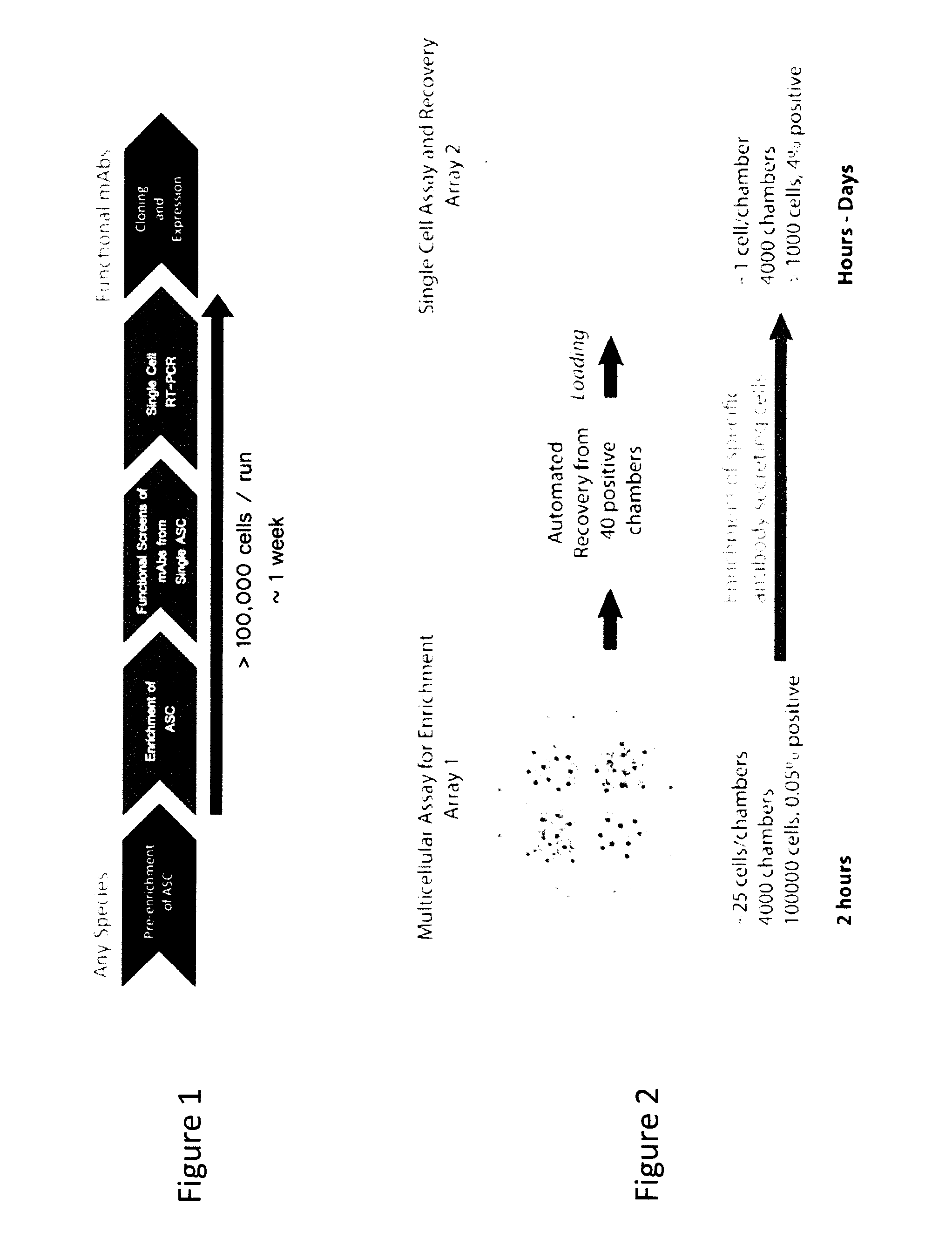
The discovery of antibodies with optimal therapeutic properties, and in particular antibodies that target surface receptors, remains a serious bottleneck in drug development.
However, even if an ASC is analyzed individually, and not within a bulk population of cells, because a single ASC generates only a minute amount of antibody, when analyzed in the volume of conventional assay formats, the antibody is too dilute, making it completely undetectable.

Method used


Image

  • Microfluidic Devices and Methods for Use Thereof in Multicellular Assays of Secretion
  • Microfluidic Devices and Methods for Use Thereof in Multicellular Assays of Secretion
  • Microfluidic Devices and Methods for Use Thereof in Multicellular Assays of Secretion

Examples


example 1

Fabrication of Microfluidic Devices

[0507]Microfluidic devices were fabricated using the protocol described in Lecault et al. (2011). Nature Methods 8, pp. 581-586 or an adapted version of this protocol (e.g., modified baking times or curing protocols) to obtain multiple layers including, from the bottom glass slide to the top: a flow chambers, a control layer, a membrane, and bath layer (FIG. 27). A cross-section of a device and a corresponding 3D-schematic of the chambers, flow channels and control channels are shown in FIG. 61 and FIG. 62, respectively. As described herein, where a control channel crosses a flow channel, a pinch valve is formed, and closed by pressurizing the control channel. A cover layer was added in certain instances to close the bath, or left omitted for an open bath such as the one shown in the device on FIG. 63.

example 2

Microfluidic Device for Cell Enrichment by Selection and Reinjection

[0508]The microfluidic device shown in FIG. 63 was used to implement an effector cell (antibody secreting cell) selection assay with reinjection capabilities for enrichment. As shown in FIG. 64, the device includes six inlet ports 1030, six control ports 1031 for controlling each inlet, four reinjection ports, one of which is 1032 and a single output port 1033. The device contains an array of 8192 identical unit cells, one of which is depicted in FIG. 2B and FIG. 61. Each unit cell contains a chamber which is 100 μm in width, 160 μm in length and 160 μm in height. Each chamber has a volume of about 2.6 nL. The device is divided into four sub-arrays, one of which is indicated by 1032 (FIG. 64). Each sub-array contains 2048 of the 8192 total unit cells. Each sub-array has its own sub-array valve 1034, which allows each sub-array to be fluidically addressed independently of the other sub-arrays. Each sub-array has a si...

example 3

Microfluidic Device for Segregation of Effector and Readout Cells

[0509]A microfluidic device was designed to permit readout cells to be kept in separate, isolated chambers from effector cells and to allow the contents of these chambers to be mixed on demand when needed. This configuration is used to implement effector cell assays, for example, cytokine neutralization assays. The architecture is particularly useful to prevent an effector cell's secretion product (e.g., antibody) from being completely washed away when the chamber comprising the effector cell(s) is perfused with a fluid, for example, when provided with fresh culture medium to maintain viability over extended periods of time. This configuration is also amenable if it is useful to limit the time during which the effector cell(s) is exposed to the medium conditions required for the extracellular effect assay (e.g., when a toxic cytokine is used as an accessory particle).

[0510]As shown in FIG. 65, this device consists of s...


PUM

PropertyMeasurementUnit
diameteraaaaaaaaaa
timeaaaaaaaaaa
time-aaaaaaaaaa

Abstract

Methods and devices are provided herein for identifying a cell population comprising an effector cell that exerts an extracellular effect. In one embodiment the method comprises retaining in a microreactor a cell population comprising one or more effector cells, wherein the contents of the microreactor further comprise a readout particle population comprising one or more readout particles, incubating the cell population and the readout particle population within the microreactor, assaying the cell population for the presence of the extracellular effect, wherein the readout particle population or subpopulation thereof provides a direct or indirect readout of the extracellular effect, and determining, based on the results of the assaying step, whether one or more effector cells within the cell population exerts the extracellular effect on the readout particle. If an extracellular effect is measured, the cell population is recovered for further analysis to determine the cell or cells responsible for the effect.

Description

CROSS REFERENCE TO RELATED APPLICATION[0001]This application claims priority from U.S. Provisional Application Ser. No. 61 / 806,329, filed Mar. 28, 2013, the disclosure of which is incorporated by reference in its entirety for all purposes.BACKGROUND OF THE INVENTION[0002]The cell is the fundamental unit of life and no two cells are identical. For example, differences in genotype, phenotype and / or morphological property can contribute to cellular heterogeneity. Indeed, “seemingly identical” clonal populations of cells have been shown to display phenotypic differences among cells within the population. Cellular differences exist across all levels of life, ranging from bacterial cells to partially differentiated cells (for example, adult stem and progenitor cells) to highly differentiated mammalian cells (for example, immune cells). Differences in cellular state, function and responses can arise from a variety of mechanisms including different histories, different differentiation state...

Claims


Application Information

Patent Timeline
Patent Type & Authority Applications(United States)
IPC IPC(8): G01N33/50C12Q1/68G01N35/00C12N5/0781C12N5/078B01L3/00G01N33/68
CPCG01N33/5052B01L2200/0647B01L3/502715B01L3/502761G01N33/5047C12Q1/6888G01N33/6845C12N5/0635C12N5/0634G01N35/0099B01L3/50273G01N33/6854B01L2300/0838B01L2300/0627B01L2300/14B01L2200/12B01L2300/0887B01L2300/0877B01L2300/04B01L3/502738G01N33/505G01N33/54313C07K16/00C07K16/1018C07K16/40C07K2317/14C07K2317/21C07K2317/70G01N33/5005
Inventor RICICOVA, MARKETAHEYRIES, KEVIN ALBERTZAHN, HANSPETRIV, OLEHLECAULT, VERONIQUESINGHAL, ANUPAMDA COSTA, DANIEL J.HANSEN, CARL L. G.NELSON, BRADNIELSEN, JULIELISAINGO, KATHLEEN
Owner THE UNIV OF BRITISH COLUMBIA
Why Eureka
Eureka Blog
Learn More