
Biological fluid filtration assembly

a technology of biological fluid and assembly, which is applied in the direction of separation process, laboratory glassware, instruments, etc., can solve the problems of poor prognosis, high recurrence rate of bladder cancer, and the most expensive cancer to trea

Inactive Publication Date: 2016-08-04
CANCER RES TECH LTD
View PDF3 Cites 14 Cited by
  • Summary
  • Abstract
  • Description
  • Claims
  • Application Information

AI Technical Summary

Benefits of technology

The present invention is about creating a convenient and efficient way to collect and store biological material from bodily fluids. This can be useful for patients and medical professionals in diagnosing and monitoring conditions. The invention includes easy-to-use filtration assemblies for collecting the material and methods for its storage. This can provide significant benefits to patients as they can have the material collected from them and stored for future analysis by an analyst or medical practitioner, or mailed to a testing facility for analysis. Overall, the invention offers a simple and effective way to capture and store biological material for various medical purposes.

Problems solved by technology

However, the recurrence rate for these tumours is very high, with around 70% of the patients experiencing relapses, and up to 25% of these recurrences will progress to muscle invasive cancers (MIBC; stage T2-4) with a poor prognosis.
The high recurrence rate and the risk of progression require life-long surveillance with periodic cystoscopy, making bladder cancer the most expensive cancer to treat (Avritscher et al., 2006).
As less than 10% of all patients presenting with microscopic or visible hematuria will be diagnosed with bladder cancer, the number of cystoscopies performed to rule out bladder cancer is high and places a considerable burden on the healthcare system.
Moreover, as cystoscopy is an invasive method that causes considerable discomfort to the patients, there is an unmet need for noninvasive techniques for reliable and cost-effective diagnosis and surveillance of bladder cancer.
However, it has a low sensitivity for detection of NMIBC (10-20%).
To date, none of these tests has achieved widespread use in clinical practice due to low specificity (Liou, L. S. (2006).
One of the main challenges when using urinary DNA markers for diagnosis and surveillance of bladder cancer is to obtain a sufficient number of cells for downstream analysis.
This limits the usefulness of urinary DNA markers in the non-invasive detection and monitoring of disease and disease progression.

Method used


Image

  • Biological fluid filtration assembly
  • Biological fluid filtration assembly
  • Biological fluid filtration assembly

Examples


examples

[0105]The following examples are set forth so as to provide those of ordinary skill in the art with a complete disclosure and description of how to practise the invention, and are not intended to limit the scope of the invention.

Capture of Cells on Micromembrane Filters

[0106]The following demonstrates the utility of membrane filters for the capturing of cells from urine for analysis.

Collection of Samples

[0107]Voided morning urine samples were collected from bladder cancer patients admitted for cystoscopy and transurethral resectioning (TURBT) at Herlev Hospital, Denmark and from healthy volunteers without known urological malignancies. Samples were sent to the Danish Cancer Research Center where they were processed within 4-6 hours after collection.

Processing of Urine Samples

[0108]For all patients and controls, 50 ml from each urine sample was sedimented by centrifugation, 2000×g for 10 min, the pellet was washed in PBS followed by another 10 min centrifugation. The supernatant was ...


PUM

PropertyMeasurementUnit
volumeaaaaaaaaaa
lengthaaaaaaaaaa
lengthaaaaaaaaaa

Abstract

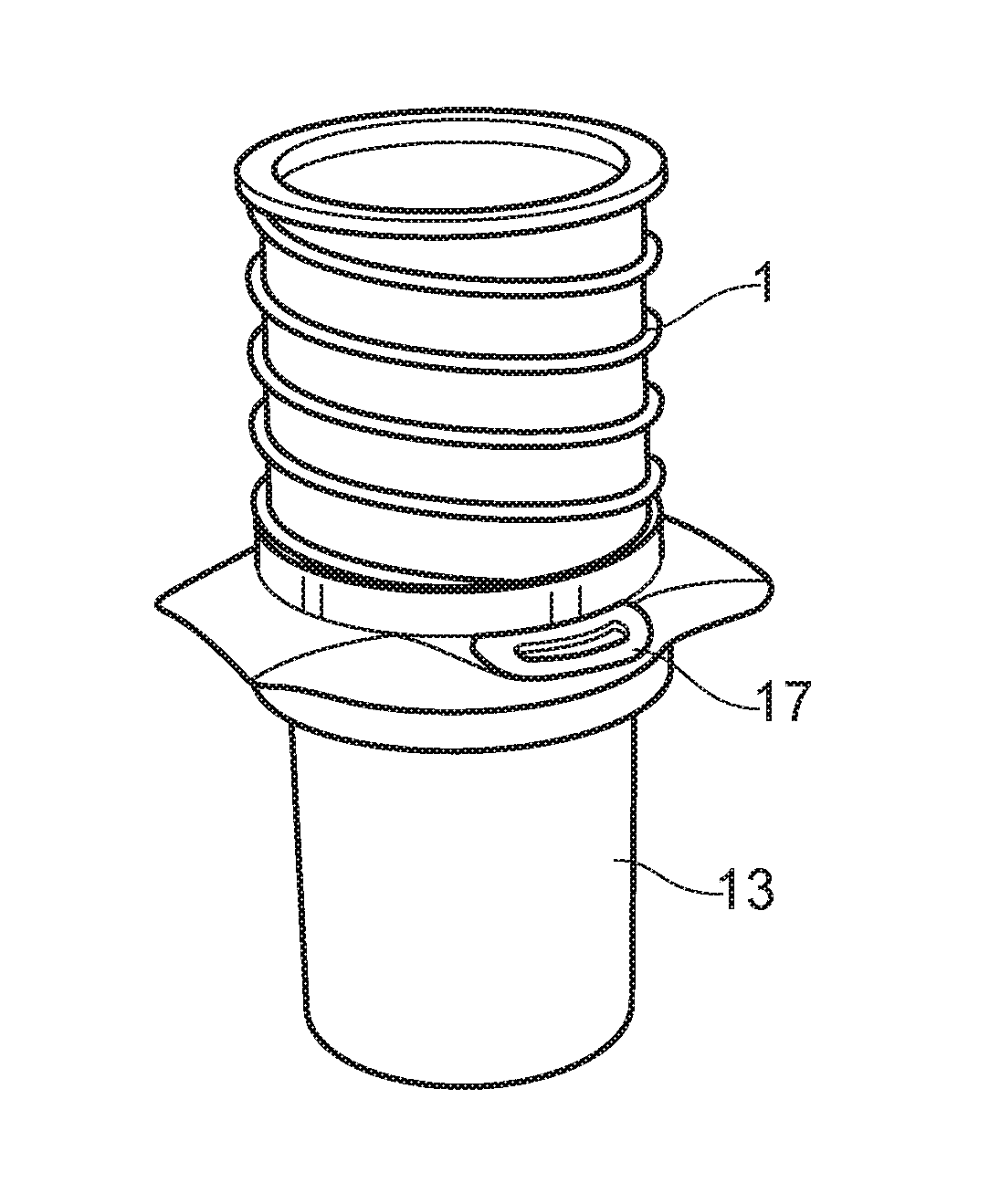
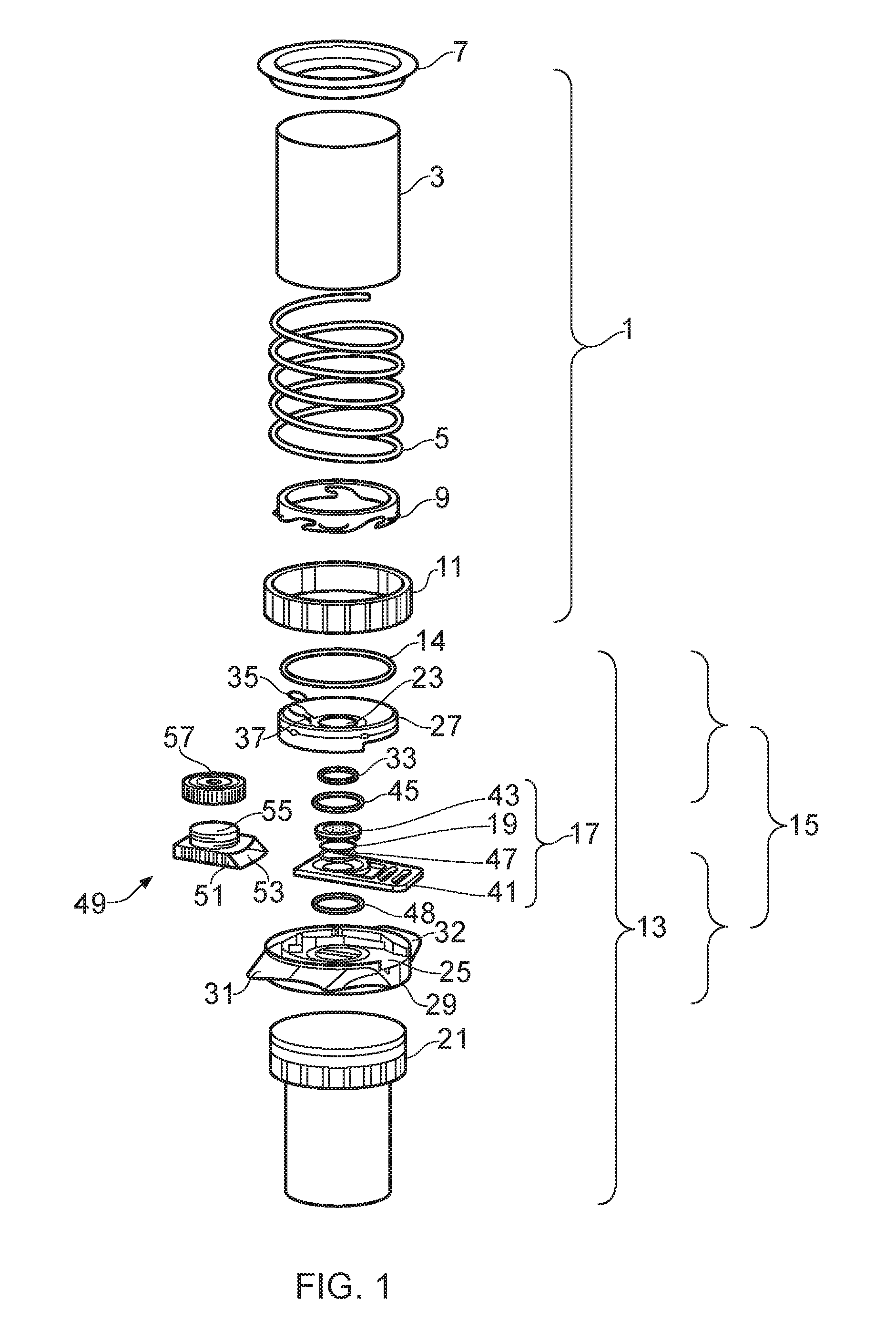
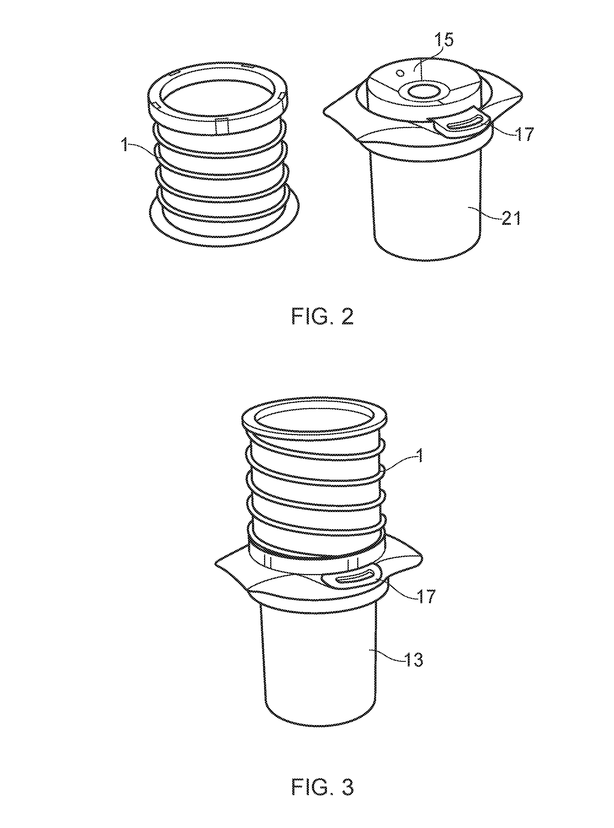
The present invention relates to biological fluid filtration assemblies and to methods of using such assemblies. The biological fluid filtration assembly has a filtration device for filtering a biological fluid sample and a storage unit, the storage unit having a body configured to engage with a removable filter cartridge of the device such that, when engaged, the filter of the filter cartridge is sealed within the body of the storage unit.

Description

[0001]This application claims priority benefit of GB1316347.2 which was filed 13 Sep. 2014 and is incorporated herein in its entirety.FIELD OF THE INVENTION[0002]The present invention relates to biological fluid filtration assemblies and to methods of using such assemblies.BACKGROUND OF THE INVENTION[0003]Bladder cancer is the sixth most common cancer in the world. The symptoms include microscopic or macroscopic hematuria, painful urination and polyuria; however, none of these symptoms is specific for the disease. The gold standard for diagnosing bladder cancer is cystoscopy and subsequent transurethral resection of the bladder tumour (TURBT). The sensitivity of cystoscopy for non-muscle invasive bladder cancer (NMIBC; stage Ta, Tl and Tis) is around 80% with white-light cystoscopy and >95% with fluorescence (hexaminolevulinate)-guided cystoscopy.[0004]The majority of bladder tumour patients (70-80%) are diagnosed with NMIBC, which has a relatively good prognosis. However, the re...

Claims


Application Information

Patent Timeline
Patent Type & Authority Applications(United States)
IPC IPC(8): G01N1/40B01D27/08C12Q1/68B01L3/00A61B10/00C12N15/10
CPCB01L3/502C12Q1/6886B01L2300/0832B01L2400/0487G01N1/4077A61B10/007A61B10/0096B01L2300/0681G01N2001/4088B01L2300/087B01L2300/041B01L2200/0631B01D27/08C12Q2600/154C12Q2600/158C12Q1/6806C12N15/1017B01L2400/0478B01L2300/12B01L2200/04
Inventor GULDBERG, PERSTEVEN, KENNETH ERIC
Owner CANCER RES TECH LTD
Learn More