One-photon integrated neurophotonic systems

a neurophotonic system and integrated technology, applied in the field of tissue functional imaging apparatus and method, can solve the problems of not being able to record electrically from specific cell types, still very far from understanding how large ensembles of neurons in the brain interact to process information, and not being able to understand the interaction between large ensembles of neurons in the brain to process information, etc., to achieve high throughput screening and accelerate drug discovery

Inactive Publication Date: 2016-06-02
THE TRUSTEES OF COLUMBIA UNIV IN THE CITY OF NEW YORK +2
View PDF2 Cites 77 Cited by
  • Summary
  • Abstract
  • Description
  • Claims
  • Application Information

AI Technical Summary

Benefits of technology

[0006]In some aspects, a new approach of integrated neurophotonics is provided. Integrated neurophotonics is a novel paradigm for functional optical imaging that surmounts the limits of present methods. It permits functional imaging with cellular resolution in highly scattering brain tissue, can offer complete coverage of all neurons within target volumes, and has eventual prospects for human applications. This approach is based on distributing a dense 3-D lattice of emitter and detector pixels within the brain itself, spaced by distances on the order or less than the optical attenuation length. These pixel arrays are embedded onto neurophotonic probes, realized as implantable, ultra narrow shanks that leverage recent advances in nanoprobe-based electrophysiology and integrated nanophotonics. Used with functional optical reporters, one 25-shank probe module is capable of recording acti...

Problems solved by technology

However, we are still very far from understanding how large ensembles of neurons in the brain interact to process information.
It is not possible, though, to record electrically from specific cell types, and up-scaling recording density to track the activity of every neuron in an extended brain region appears infeasi...

Method used


Image

  • One-photon integrated neurophotonic systems
  • One-photon integrated neurophotonic systems
  • One-photon integrated neurophotonic systems

Examples


example 1

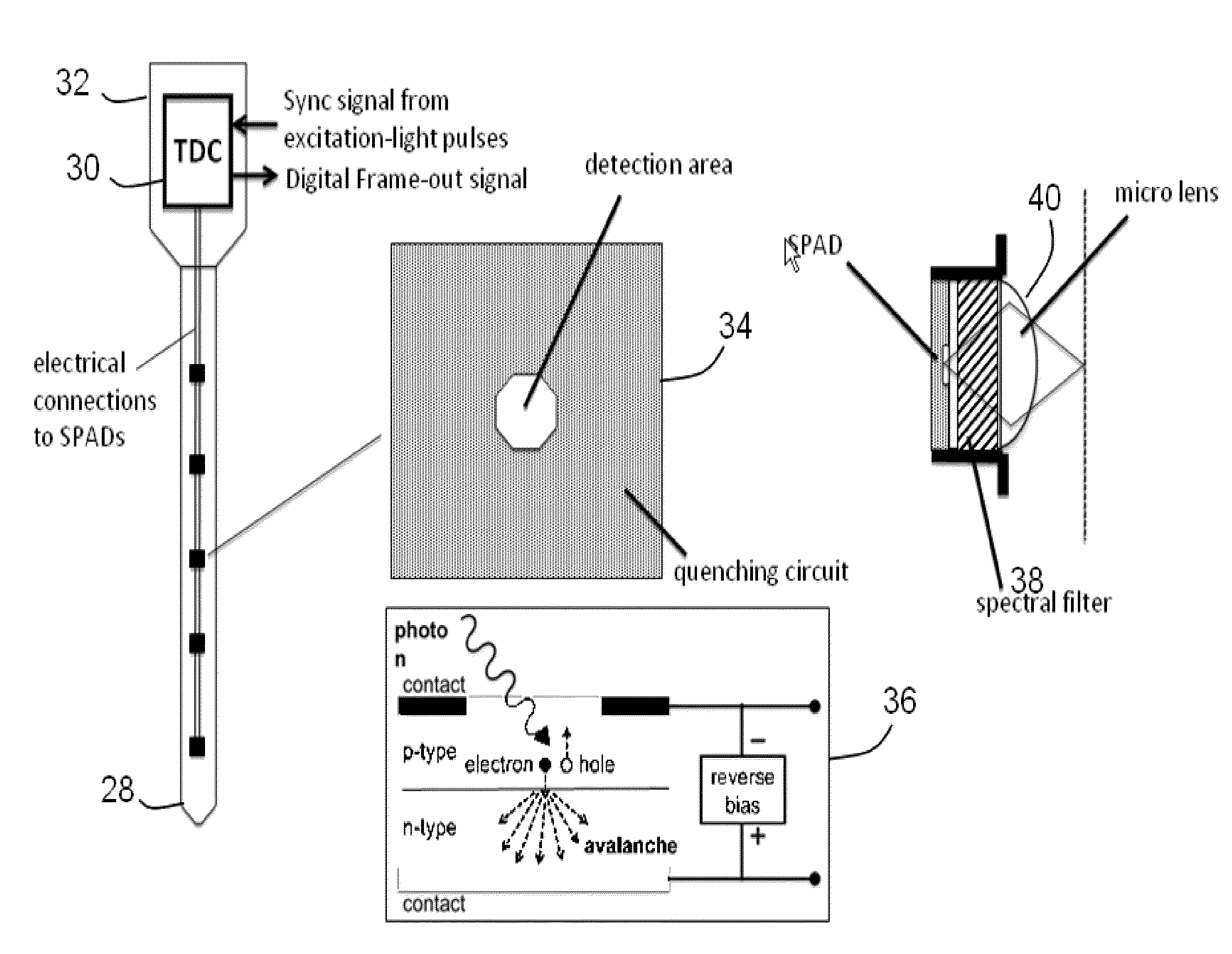
[0097]A simulation based on the prototype design for the neurophotonic probe arrays has been executed (FIG. 8). This demonstrates that the integrated neurophotonic probes described herein are capable of recording from very large populations of neurons, and can provide single-cell resolution. This modeling also shows that it is possible to assign calcium events that contribute the resulting, very-high-dimensional data stream to specific neurons. To validate the paradigm for optical spike sorting, a simulation involving 360 neurons randomly in a 282×282×50 μm3 unit volume (0.004 mm3) has been carried out. This mimics cell densities that are observed via two-photon imaging from the mouse visual cortex in vivo. The excitation intensity provided by each E-pixel to each neuron was determined, and the collection efficiency from each neuron to each D-pixel within one attenuation length was numerically calculated. Each specific combination of emitters and detectors yields an independent meas...


PUM


Abstract

An apparatus and method for detecting functional cellular activity within a volume of a tissue. The method includes inserting a three-dimensional array of optical emitters and optical detectors into a volume of a tissue, where the tissue volume includes one or more cells labeled with an optical reporter of cellular activity; illuminating the one or more cells with photons from the optical emitters of the three-dimensional array to generate optical signals from the optical reporter that labels the one or more cells; and detecting the optical signals using the optical detectors of the three-dimensional array, where the illumination includes one-photon excitation of the optical reporter.

Description

BACKGROUND[0001]1. Field of the Invention[0002]The invention relates to an apparatus and method for functional imaging of tissue.[0003]2. Related Art[0004]Over the past few decades, our understanding of the properties of individual neurons and their role in brain computations has advanced significantly. However, we are still very far from understanding how large ensembles of neurons in the brain interact to process information. For monitoring neuronal activity, extracellular electrical recording provides unparalleled temporal resolution. It is not possible, though, to record electrically from specific cell types, and up-scaling recording density to track the activity of every neuron in an extended brain region appears infeasible. Functional imaging by free-space two-photon microscopy enables single-cell resolution of large neuron ensembles at anatomical densities and provides cell-type specificity of activity via genetically encoded fluorescent reporters. But it works ideally only w...

Claims


Application Information

Patent Timeline
IPC IPC(8): A61B5/00
CPCA61B5/0071A61B5/4029A61B5/4064A61B5/0084A61B5/685A61B5/6868A61B5/7225A61B2562/0233A61B2562/028A61B2562/0285A61B2562/046
Inventor ROUKES, MICHAEL LEECOTTON, RONALD JAMESMOREAUX, LAURENTSHEPARD, KENNETHSIAPAS, ATHANASSIOSTOLIAS, ANDREAS
Owner THE TRUSTEES OF COLUMBIA UNIV IN THE CITY OF NEW YORK
Why Eureka
Try Eureka