Systems and methods for single cell isolation and analysis

Inactive Publication Date: 2016-03-10
RGT UNIV OF MICHIGAN
View PDF3 Cites 11 Cited by
  • Summary
  • Abstract
  • Description
  • Claims
  • Application Information

AI Technical Summary

Benefits of technology

The present patent relates to devices, systems, and methods for isolating and analyzing single cells. The invention provides a system for capturing single cells with a high efficiency, even at low cell numbers. The system includes a cell capture device with multiple wells that capture cells based on their adhesion to surfaces, and a fluid transport device that prevents cell damage during transport. The system can also include a microscope for observing cells and sensors for pH, oxygen, glucose, proteins, and metabolic byproducts. The invention also provides a cell culture device with multiple chambers for adherent and suspension culture, as well as interaction channels for cell-cell interactions. The method involves culturing cells in the adherent and suspension chambers, and utilizing the interaction channels for analysis. The invention allows for the isolation and analysis of cancer stem cells and fibroblasts, and the study of cell-cell interactions.

Problems solved by technology

Cancer cell heterogeneity is one of key challenges in modern cancer study.
These mechanisms cannot be easily studied by conventional dish-based assays.
However, these methods lack the ability to retrieve a target single cell for further analysis (e.g., genotyping) and assays (e.g., drug-screening).
However, detaching cells from the special film limits spatial resolution and it is difficult to handle cells over the film.
On top of that, the cell detachment based on photodegradation of the substrate film, which generates acid, may lead to toxicity to the cells (Kimio et al., Proceedings of MicroTAS 2013 100-102).
However, cell viability was poor because of heat-induced cell necrosis under direct laser irradiation.
Recently, cell detachment using ultrasound-induced cavitation was demonstrated (Baac et al., (2012) Sci. Rep. 2, 989), but unfortunately this approach only works on Petri dishes and is not compatible with microfluidic arrangement due to acoustic attenuation by PDMS.

Method used


Image

  • Systems and methods for single cell isolation and analysis
  • Systems and methods for single cell isolation and analysis
  • Systems and methods for single cell isolation and analysis

Examples


example 1

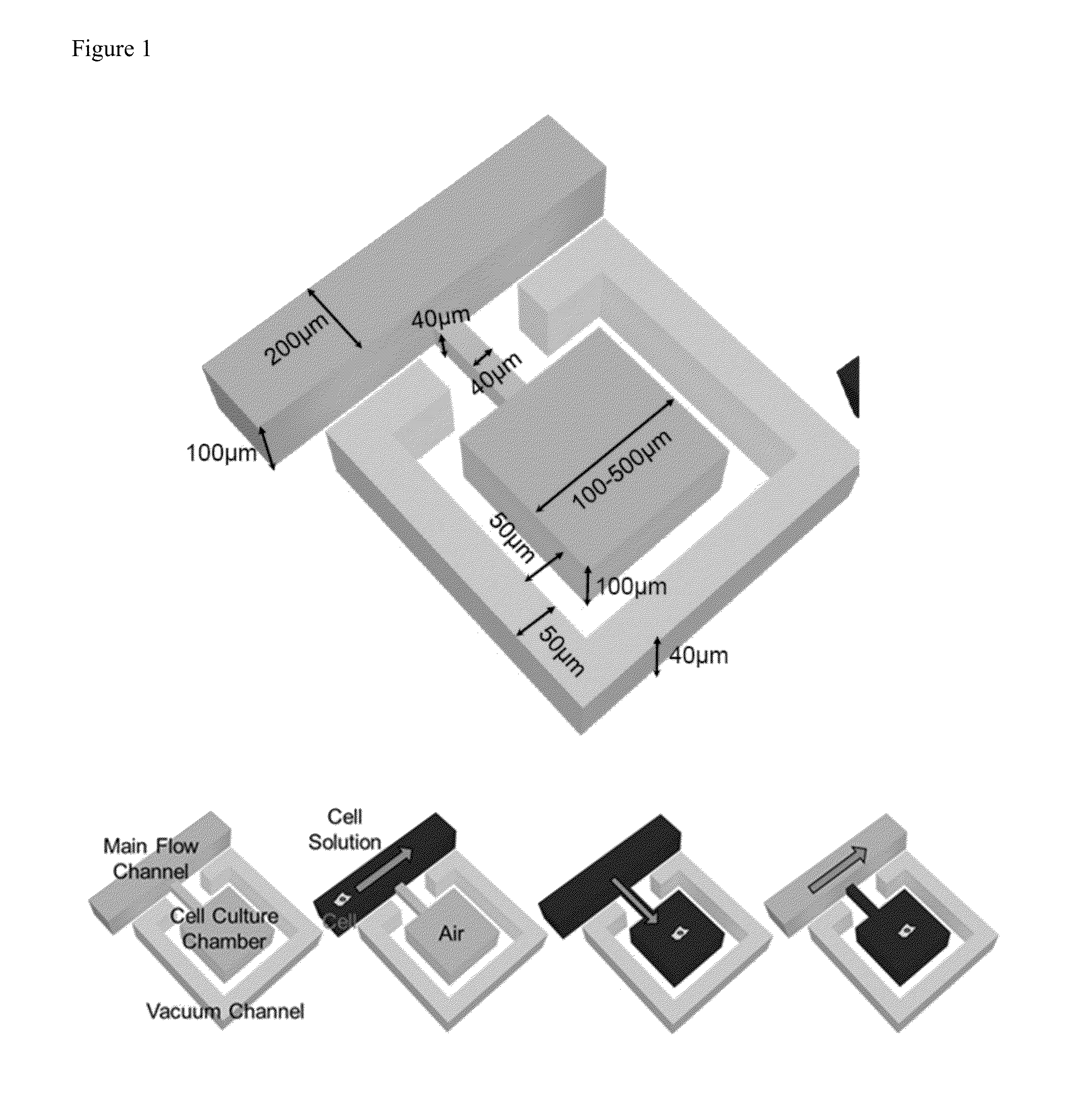
[0092]Operation of the Small Sample Capture Device

[0093]The device eliminates dead volume by driving all of the cell solution into the microfluidics. FIG. 1 shows the operation of a single capture well. The design composes of a main-channel, which transports the cell solution, a cell culture chamber and vacuum channel, which drives the solution into the chamber. One useful characteristic of PDMS is its gas permeability, so the air can diffuse through the PDMS sidewall to the vacuum channel. As a result, the cell can be loaded into the chamber with the cell solution. FIG. 2 demonstrates a cell loading process. One cell was loaded into the chamber.

[0094]FIG. 3 illustrates the operation of the device. First, liquids are pulled downstream to drive the cell solution into the main channel. Then, the vacuum channel is sued to drive the solution and cells into the chamber. If there is any residual solution in the main channel as shown in FIG. 3(C), it is driven downstream and the process is...

example 2

[0101]Design and Fabrication

[0102]The co-culture platform is composed of an inner suspension culture chamber, an outer adherent culture chamber, and narrow interaction channels connecting them (FIG. 7(a)). The whole device comprises or consists of 120 co-culture units (15 by 8) (FIG. 7(b)), and each unit is composed of the two culture chambers connected by 7 narrow (cross-section 3 μm by 20 μm and 100 μm long) channels. To facilitate suspension and adherent culture on the same device, two layers of PDMS are used in fabrication. The bottom layer was patterned with indented microwells that were selectively coated with Poly-hydroxyethylmethacrylate (polyHEMA, Sigma-Aldrich), which has been extensively used as an adhesion blocking coating material [34]. The top channel layer is patterned with microfluidic channels for flow control and chambers for co-culture.

[0103]These two PDMS layers (channel layer and substrate layer) were separately fabricated using a soft lithography processes, and...


PUM

PropertyMeasurementUnit
Temperatureaaaaaaaaaa
Lengthaaaaaaaaaa

Abstract

The present disclosure relates to devices, systems, and methods for single cell isolation and analysis. In particular, the present disclosure relates to systems and methods for isolating single cells present at low numbers.

Description

[0001]The present application claims priority to U.S. Provisional Patent Application Ser. No. 62 / 047,819, filed Sep. 9, 2014, the disclosure of which is herein incorporated by reference in its entirety.FIELD OF THE INVENTION[0002]The present disclosure relates to devices, systems, and methods for single cell isolation and analysis. In particular, the present disclosure relates to systems and methods for isolating single cells present at low numbers.BACKGROUND OF THE INVENTION[0003]Cancer cell heterogeneity is one of key challenges in modern cancer study. Due to the genomic instability of cancer cells (Negrini et al., (2010) Nat Rev Mol Cell Biol. 11(3):220-228), certain cells may have higher capability of drug resistance, metastasis and tumorgenesis (Visvader and Lindeman G J (2008) Nat Rev Cancer. 8(10):755-768). Studying these sub-populations separately can lead to effective therapeutic targets. For example, the state transition, such as the epithelial-to-mesenchymal transition (E...

Claims


Application Information

Patent Timeline
IPC IPC(8): B01L3/00C12M1/00C12M3/06
CPCB01L3/502761C12M23/16C12M23/20B01L2200/0668B01L2300/0663B01L2300/0654B01L2400/049B01L2300/12B01L2300/0893C12M47/04B01L3/502707B01L2300/0645B01L2300/0816B01L2300/0864B01L2400/0655
Inventor YOON, EUISIKCHEN, YU-CHIH
Owner RGT UNIV OF MICHIGAN
Why Eureka
Try Eureka