
Colorectal Cell Sampling Device

Active Publication Date: 2015-08-20
ORIGIN SCI
View PDF11 Cites 8 Cited by
  • Summary
  • Abstract
  • Description
  • Claims
  • Application Information

AI Technical Summary

Benefits of technology

The device described in this patent prevents contamination and material loss during digital sampling by keeping a flexible membrane within the insertion member both on insertion and withdrawal. The device can be used for various analysis such as DNA quantitation, DNA extraction, cytological / cytochemical investigation, and biochemical tests. The device provides a sample with low levels of contaminants and high concentration of cells from the colorectal mucosal surface being sampled, which improves the accuracy of screening.

Problems solved by technology

A serious obstacle to early diagnosis of CRC is the absence of early, readily identifiable clinical manifestations in the majority of cases.
However, the late stages of the disease are also associated with invasive or metastatic tumours.
Unfortunately, there is presently no CRC screening technique that combines low invasiveness, simplicity and low cost with high sensitivity and specificity.
However, both of these methods have significant drawbacks.
Flexible colonoscopy / sigmoidoscopy is regarded as a precise and reliable diagnostic procedure, however, its invasiveness, cost and requirement for skilled and experienced specialists to carry out the procedure make its use in routine screening impractical.
FOBT is cheap and simple, however, it produces unacceptably high rates of both false negative and false positive results.
However, the method of obtaining these samples (an invasive colonic lavage procedure) suffered from the same disadvantages as sigmoidoscopy / colonoscopy, and required detailed cytological analysis of the sample once obtained.
However, these ambitious claims have generated substantial doubts due to the low likelihood of the presence of well-preserved colonocytes in an aggressive anaerobic environment such as that found in the faeces.
Furthermore, morphological evidence presented in their 1991 reference was unconvincing.
S84-S93], but have not produced any practical advances based on the outcomes of their studies.
However, despite the lack of practical advances by P. P. Nair and his colleagues, the use of human stool for diagnostic and research purposes remains an active research area as it is not associated with any invasive intervention.
Whilst DNA directly isolated from homogenized faeces can be amplified and analysed for the presence of cancer-associated genetic alterations, the absence of a highly reliable single molecular biomarker for cancer resulted in the use of multiple molecular markers reflecting a number of genetic alterations known to be present in malignant cells at relatively high frequencies.
Although detection of colorectal tumours by multi-target molecular assays appears to be feasible, the validity of these methods for screening purposes remains questionable due to the high cost and relative complexity of laboratory procedures involved.
It is, however, apparent that homogenized stool is a difficult material for human DNA extraction.
In particular, the abundance of bacteria in faeces can interfere with colonocyte DNA recovery procedures, and rapid mammalian DNA damage and degradation occur in the presence of anaerobic bacterial flora of the human colon.
The development of approaches based upon exfoliated colonocyte isolation has been slow partially due to a surprising lack of knowledge on cell exfoliation in the gut both in normal physiological conditions and in disease.
): A463] or DNA [Villa, E. et al., Gastroenterology 1996; 110: 1346-1353] in dispersed or homogenized stool samples obtained from CRC patients, however the differences between healthy people and cancer patients observed in those studies were not large enough to be considered diagnostically valid.
Although the technique and results of its initial trials apparently highlighted a very efficient, simple and inexpensive approach to CRC screening, it had a number of substantial faults (apparent difficulties of whole stool handling and especially impossibility of the procedure standardization) preventing its commercialization and serious introduction into clinical practice.
These problems, difficulty of standardization being the crucial one, cause serious doubts with regard to using exfoliated colonocytes isolated from stool samples for wide scale CRC screening.
However, although one can achieve a contact with the rectal mucocellular layer by employing this simple manipulation, significant losses of material and simultaneous contamination with irrelevant squamous epithelium of the anal canal are inevitable during the removal of the finger from the rectum.
Smears prepared from gloves used for rectal examination have shown well-preserved colonocytes, combined with a high level of contamination by cells of the squamous epithelium.

Method used


Image

  • Colorectal Cell Sampling Device
  • Colorectal Cell Sampling Device
  • Colorectal Cell Sampling Device

Examples


Embodiment Construction

Description of Cell Sampling Embodiments

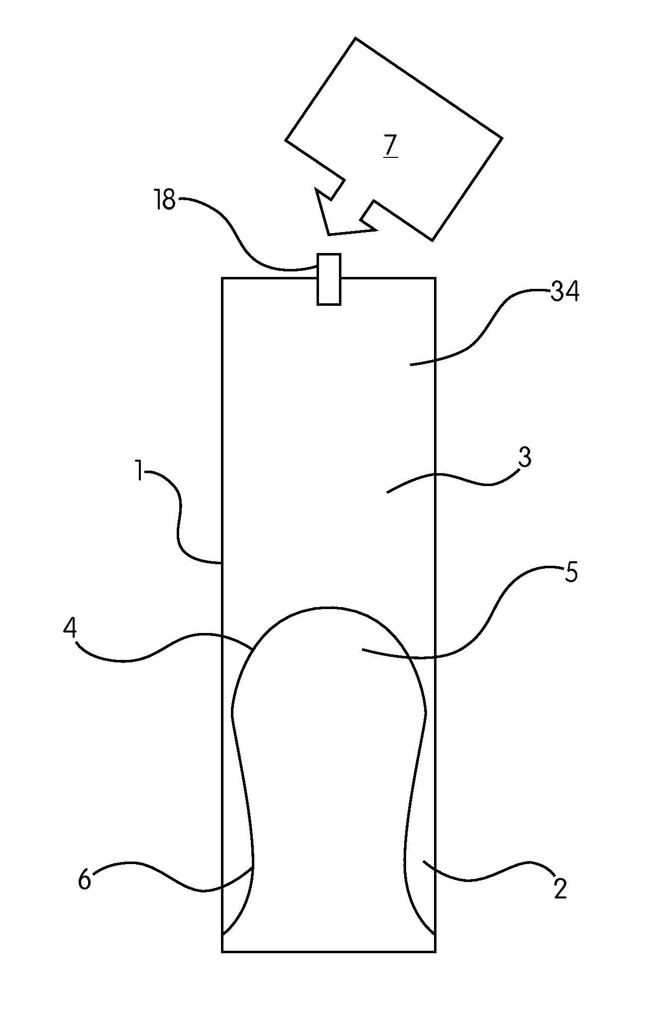
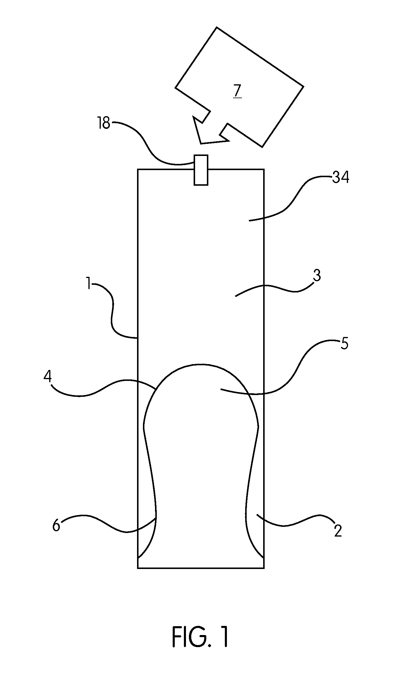
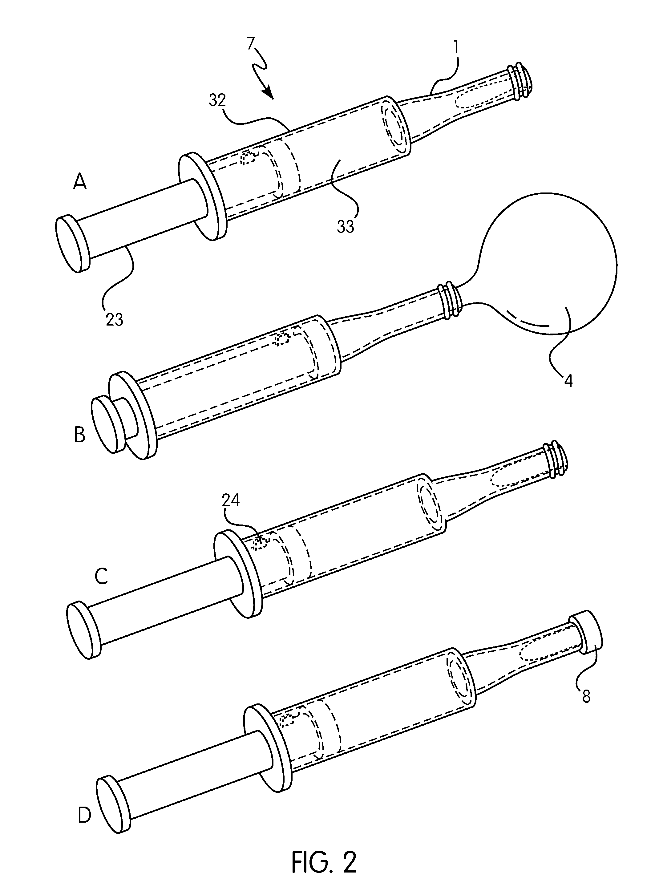
[0128]The cell sampling device of FIG. 1 is designed for insertion into a rectal cavity. The device comprises a substantially cylindrical insertion member 1 with an interior cavity 3, closed at the distal insertion end 2 by a flexible and resilient membrane 4 which is sealingly attached to the member 1 at the distal end 2. In the position shown in FIG. 1, the membrane 4 is held within the cavity 3, and is adapted to emit from the cavity 3 when the cavity 3 is pressurised by means 7 (shown in more detail in FIG. 2). The membrane 4 has a cell sampling surface 5 which in the rest position shown in FIG. 1 is the inner surface, but when the membrane emits is the outer surface, and an opposing surface 6 which in the rest position is the outer surface, but which becomes the inner surface when the membrane emits. The membrane is made of nitrile, latex or a rubber based substance. At the proximal end 34, the cavity 3 is closed by a self-sealing valve 1...


PUM


Abstract

The present invention relates to a device for collecting a sample of exfoliated cells from a colorectal mucosal surface of a human subject which comprises a colorectal insertion member (1) having a distal, insertion end (2), a proximal end (34) and a closable interior cavity (3), a flexible membrane having an outer, cell sampling surface (5) and an inner surface, wherein said membrane is sealingly attached to the distal, insertion end of said insertion member and held within the interior cavity, such that, in use, pressurization of the interior cavity to at least a first elevated pressure causes the membrane to emit from the distal end of said insertion member to make contact with the colorectal mucosal surface and pressurization of the interior cavity to a second reduced pressure causes the membrane to invert and return to the interior cavity of said insertion member. A kit comprising said device and methods of colorectal cell sampling using said device are also described.

Description

FIELD OF THE INVENTION[0001]The present invention relates to a device for collecting a sample of exfoliated cells from a colorectal mucosal surface of a human subject, to a kit comprising said device and to methods of colorectal cell sampling using said device.BACKGROUND OF THE INVENTION[0002]Sporadic colorectal cancer (CRC) is one of the most frequently occurring and deadly of the oncological diseases affecting people in developed Western countries. It predominantly affects people over the age of 50.[0003]A serious obstacle to early diagnosis of CRC is the absence of early, readily identifiable clinical manifestations in the majority of cases. It is only in the advanced stages of the disease, when larger tumours have formed, resulting in pain, bleeding and symptoms of obstruction, that the disease is readily diagnosed. However, the late stages of the disease are also associated with invasive or metastatic tumours. Thus, detection of colorectal tumours prior to the advanced stages o...

Claims


Application Information

Patent Timeline
IPC IPC(8): A61B10/04A61B10/00
CPCA61B10/04A61B2010/0208A61B10/0096A61B10/02A61B10/0038
Inventor LOKTIONOV, ALEXANDRELLEWELYN, ANDREW HUMPHREYLYWOOD, RUPERT CHARLES GIFFORDBANDALETOVA, TATIANAFERRETT, COLIN GEORGELYWOOD, HUGO GEOFFREY GIFFORD
Owner ORIGIN SCI
Learn More