
System and method for non-invasive patient-image registration

Active Publication Date: 2015-02-19
KOREA INST OF SCI & TECH
View PDF19 Cites 16 Cited by
  • Summary
  • Abstract
  • Description
  • Claims
  • Application Information

AI Technical Summary

Benefits of technology

The system and method described in this patent allow for non-invasive registration between a patient and a 3D medical image using near infrared light and a blood vessel model. This eliminates the need for fiducial markers and reduces skin deformation and registration errors compared to conventional methods. The position registration is based on the feature point of a blood vessel pattern distributed throughout the body, making it applicable to various surgical areas and medical images. Additionally, a center line is extracted from each image and the branch point of each skeleton line is selected as the feature point to further reduce registration errors.

Problems solved by technology

However, in the minimally invasive surgery, an operating surgeon cannot directly see an affected area and therefore indirectly checks an image gained from the endoscope through a monitor.
At this time, it is not easy to observe a targeted affected area because a view direction of the surgeon is different from a direction of the endoscope and the endoscope is short-sighted.
Further, it is unintuitive and thus difficult to treat the affected area by controlling a surgical instrument while watching an image from the endoscope.
In this surgery method, an operating surgeon cannot directly see an affected area and also cannot use the image from the endoscope in real time.
On the other hand, the non-invasive method is not applicable to microsurgical operation due to low precision.
However, the registration between the 2D sectional image and the 3D medical image needs long calculation time and is vulnerable to soft tissue transformation of a human body.
Also, the registration between the 3D scan model of a patient's skin surface and a skin surface model extracted from the 3D medical image may have a large error due to change in shape of the skin surface.

Method used


Image

  • System and method for non-invasive patient-image registration
  • System and method for non-invasive patient-image registration
  • System and method for non-invasive patient-image registration

Examples


Embodiment Construction

[0037]Hereinafter, exemplary embodiments according to the present invention will be described in detail with reference to accompanying drawings. Prior to this, terms or words used in this specification and claims have to be interpreted as the meaning and concept adaptive to the technical idea of the present invention rather than typical or dictionary interpretation on a principle that an inventor is allowed to properly define the concept of the terms in order to explain his / her own invention in the best way.

[0038]Therefore, because embodiments disclosed in this specification and configurations illustrated in the drawings are nothing but preferred examples of the present invention and do not fully describe the technical idea of the present invention, it will be appreciated that there are various equivalents and alterations replacing them at the filing date of the present application.

[0039]In minimally invasive surgery, a three-dimensional (3D) medical image (computer tomography (CT),...


PUM


Abstract

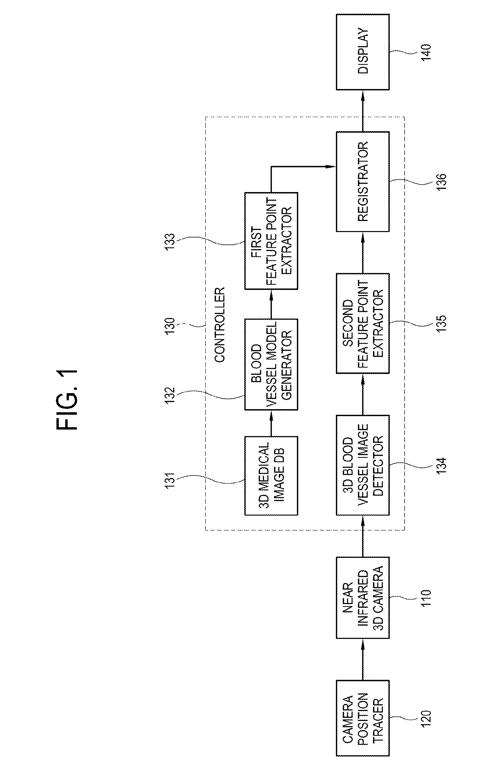
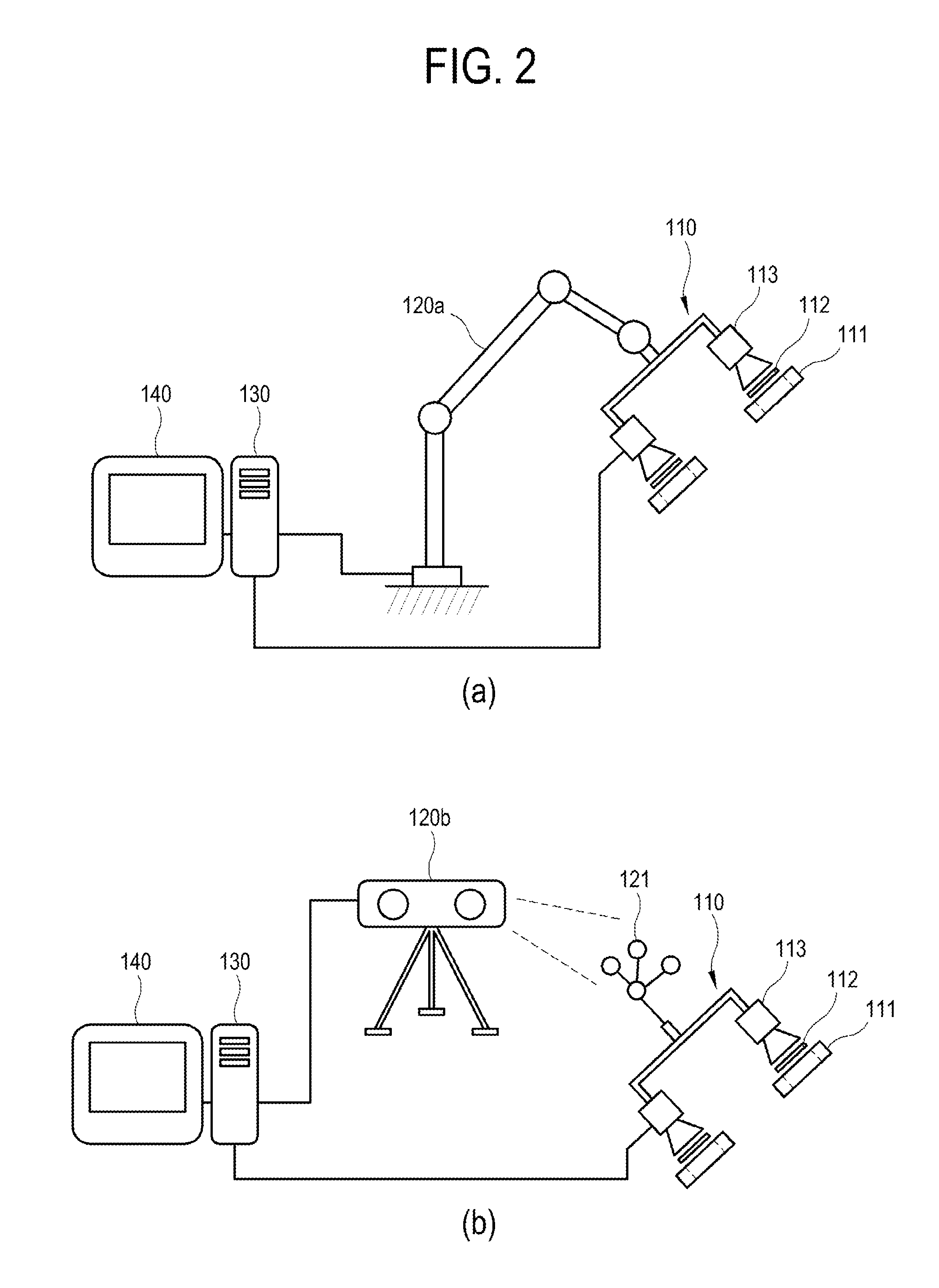
Disclosed is a system for non-invasive registration between a patient and a three-dimensional (3D) medical image, the system comprising: a near infrared 3D camera 110 which extracts a 3D blood vessel image I2 of a patient's registration target area during surgical operation; a camera position tracer 120 which traces a position of the near infrared 3D camera 110 and calculates a real world coordinate system of the 3D blood vessel image I2; a controller 130 which extracts a first blood vessel pattern from a 3D medical image I1 of the registration target area, extracts a second blood vessel pattern from the 3D blood vessel image I2, and performs position registration between the patient and the 3D medical image I1 through the extracted first and second blood vessel patterns; and a display 140 which displays a registration result calculated by the controller 130.

Description

CROSS-REFERENCE TO RELATED APPLICATION[0001]This application claims priority from Korean Patent Application No. 10-2013-0096016, filed on Aug. 13, 2013 in the Korean Intellectual Property Office, the disclosure of which is incorporated herein by reference.BACKGROUND[0002]1. Field[0003]Apparatuses and methods consistent with the exemplary embodiments relate to a system and method for non-invasive registration between a patient and a patient's three-dimensional (3D) image for registration of a position and a pose between a patient and a patient's 3D medical image, and more particularly to a system and method for non-invasive registration between a patient and a patient's three-dimensional (3D) image, which can align a patient's 3D medical image with a patient by a non-invasive method using a feature point based on a blood vessel pattern, where a patient's blood vessel is extended, without using an artificial fiducial marker.[0004]2. Description of the Related Art[0005]In general, a mi...

Claims


Application Information

Patent Timeline
IPC IPC(8): H04N13/02
CPCH04N13/0221A61B6/504A61B8/0891G06T2207/10012G06T2207/10048G06T2207/10072G06T2207/20044G06T2207/30101G06T2207/30172A61B2034/2055A61B2090/364A61B90/361G06T7/33H04N13/221A61B5/055A61B6/03A61B2576/02G16Z99/00
Inventor LEE, DEUKHEEPARK, SE HYUNGLIM, SUNGHWAN
Owner KOREA INST OF SCI & TECH
Learn More