
Portable Ultrasound System Comprising Ultrasound Front-End Directly Connected to a Mobile Device

a portable, ultrasound technology, applied in ultrasonic/sonic/infrasonic image/data processing, tomography, applications, etc., can solve the problems of limiting the use of cost and training requirements, and predominantly high cost and complexity of imaging systems

Inactive Publication Date: 2015-02-05
POCKETABONICS
View PDF11 Cites 25 Cited by
  • Summary
  • Abstract
  • Description
  • Claims
  • Application Information

AI Technical Summary

Benefits of technology

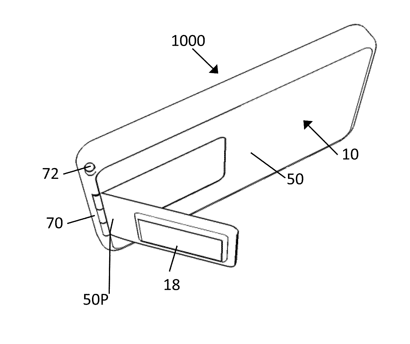
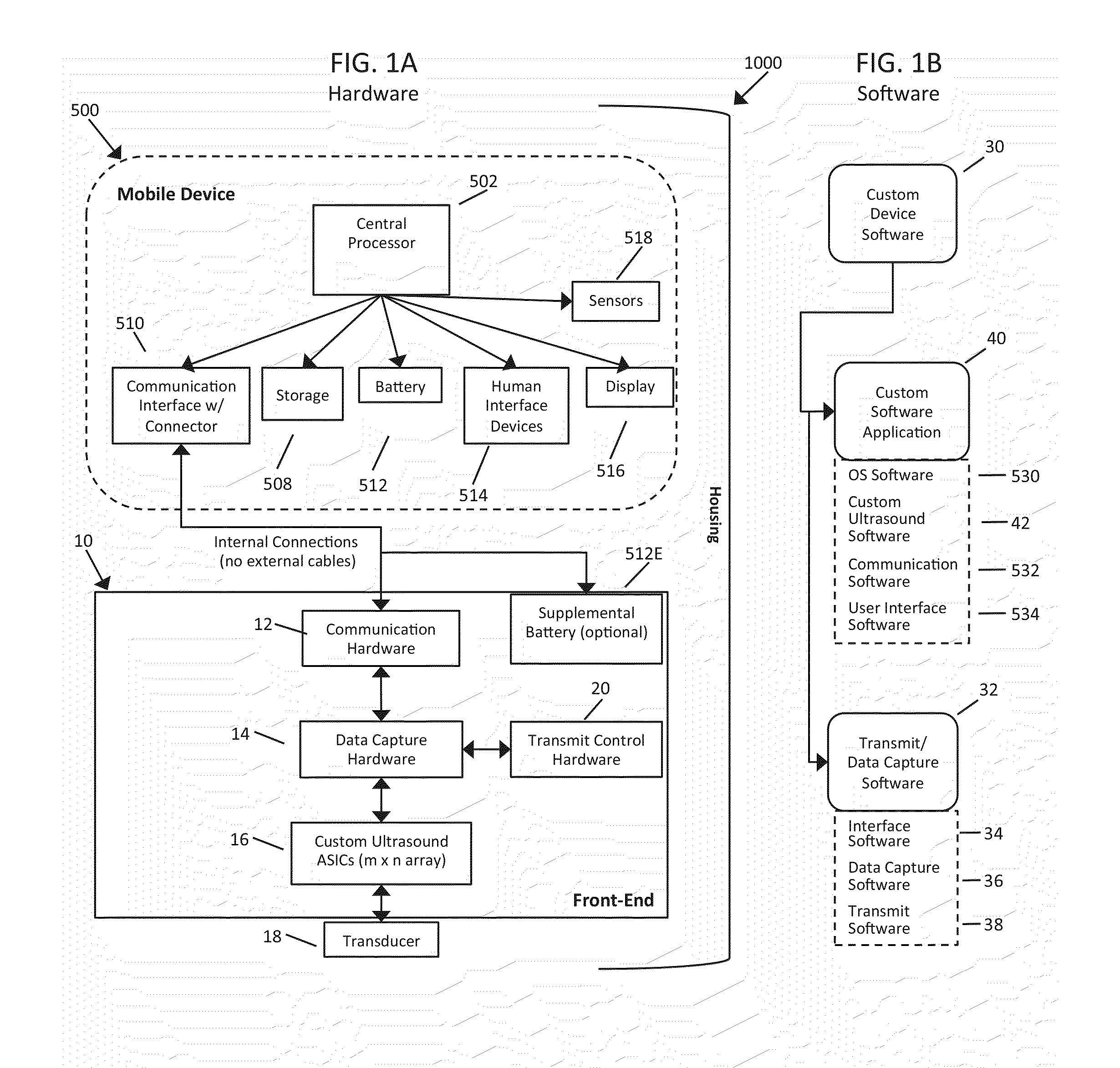
The present invention is a portable ultrasound imaging system that can be directly attached to a mobile computing device, such as a smartphone or tablet, and used for medical imaging purposes. The system includes a detachable front end component that is connected to the mobile computing device and can be rotated about one or more axes. The front end component has a movable transducer array that can be positioned over a target to be imaged and the system can be used with different imaging modes and settings. The system is designed to be easily attached and detached from the mobile computing device and can be used with different types of mobile computing devices, such as smartphones or tablets. The front end component can be sterilized and a seal can be formed with the mobile computing device to ensure a sterile environment. The front end component can be a two-dimensional ultrasound transducer or a one-dimensional ultrasound transducer. The system can also include a barrier element to shield the mobile computing device from contact with the patient when the front end component is applied to the patient. The front end component can be a separate device that is attached to the mobile computing device using a connector or a separate attachment method, such as a magnet or a clip. The front end component can be a separate device that is attached to the mobile computing device using a seal. The system can also include a locking element to fix the front end component in a desired orientation. The front end component can have a different dimensional footprint and element pitch to match the footprint and channel pitch of the mobile computing device. The system can also include a separate device for transmitting and receiving ultrasound signals. The method of using the portable ultrasound imaging system includes connecting the front end component to the mobile computing device, positioning the transducer array over the target to be imaged, selecting the desired imaging mode and settings, and activating the custom software to perform the imaging. The system can also include a barrier element to shield the mobile computing device from contact with the patient.

Problems solved by technology

Medical imaging is a field in which imaging systems are predominately very high cost and complex enough to require operation and interpretation by experienced and highly trained medical staff.
These high-tech, high-cost systems are useful for diagnostic ultrasound exams, however the cost and training requirements limit their use in many routine exams for which ultrasound can be clinical useful.
While far less expensive than high-end systems and much more portable, these systems are still fairly sophisticated and require a well-trained operator who can mentally map the image plane on the screen to the anatomy being imaged and adjust a large set of system parameters to optimize image quality.
These cables can be cumbersome for the operator and also add to the expense of the system for high channel count transducers, while at the same time making them relatively less portable.
Furthermore, the separation between the transducer and the system display requires the operator to turn his / her attention away from the patient to view the display.
This is particularly challenging during ultrasound-guided procedures.
This device is limited to making Doppler measurements, such as finding the velocity of a target object or monitoring an in utero baby's heart rate; it does not form 2D or 3D ultrasound images.

Method used


Image

  • Portable Ultrasound System Comprising Ultrasound Front-End Directly Connected to a Mobile Device
  • Portable Ultrasound System Comprising Ultrasound Front-End Directly Connected to a Mobile Device
  • Portable Ultrasound System Comprising Ultrasound Front-End Directly Connected to a Mobile Device

Examples


Embodiment Construction

[0053]Before the present systems, programming, methods and computer readable media are described, it is to be understood that this invention is not limited to particular embodiments described, as such may, of course, vary. It is also to be understood that the terminology used herein is for the purpose of describing particular embodiments only, and is not intended to be limiting, since the scope of the present invention will be limited only by the appended claims.

[0054]Where a range of values is provided, it is understood that each intervening value, to the tenth of the unit of the lower limit unless the context clearly dictates otherwise, between the upper and lower limits of that range is also specifically disclosed. Each smaller range between any stated value or intervening value in a stated range and any other stated or intervening value in that stated range is encompassed within the invention. The upper and lower limits of these smaller ranges may independently be included or ex...


PUM


Abstract

A portable ultrasound imaging system includes a mobile computing device; a detachable front end component configured for attachment to and communication with the mobile computing device, and configured to transmit and receive ultrasound signals; and programming, when installed on the mobile computing device, being executable by the mobile computing device to cause the mobile computing device to send signals to the front end component causing the front end component to transmit the ultrasound signals, and to receive signals from the front end component resulting from the front end component receiving the receive ultrasound signals, and process the receive signal and display an ultrasound image resulting from the processing. The front end component is configured to be directly joined with the mobile computing device and directly connected, without the use of an external wire or cable.

Description

FIELD OF THE INVENTION[0001]The present invention relates to the field of ultrasound imaging and is particularly well suited to the field of medical imaging. More specifically, the present invention relates to devices, methods and computer readable media for portable, ultrasound imaging.BACKGROUND OF THE INVENTION[0002]Medical imaging is a field in which imaging systems are predominately very high cost and complex enough to require operation and interpretation by experienced and highly trained medical staff. Medical ultrasound is generally considered a low cost imaging modality within the medical imaging field but utilizes imaging systems costing as much as $250K. These high-tech, high-cost systems are useful for diagnostic ultrasound exams, however the cost and training requirements limit their use in many routine exams for which ultrasound can be clinical useful.[0003]Over the past two decades a number of companies have attempted to develop low-cost, easy-to-use ultrasound systems...

Claims


Application Information

Patent Timeline
Patent Type & Authority Applications(United States)
IPC IPC(8): A61B8/00A61B8/14A61B8/08
CPCA61B8/4427A61B8/463A61B8/145A61B8/5223A61B8/4477A61B8/5207A61B8/4494A61B8/466A61B8/467A61B8/56
Inventor BLALOCK, TRAVISFULLER, MICHAELGUENTHER, DRAKEMORGAN, KARENPOMPEO, JEFF
Owner POCKETABONICS
Learn More