
X-ray imaging device and method of controlling the same

a technology of x-ray imaging and x-ray, which is applied in the field of x-ray imaging devices, can solve the problems of patient pain, patient discomfort, patient cold sensation, etc., and achieve the effect of reducing patient discomfor

Inactive Publication Date: 2014-10-02
SAMSUNG ELECTRONICS CO LTD
View PDF7 Cites 11 Cited by
  • Summary
  • Abstract
  • Description
  • Claims
  • Application Information

AI Technical Summary

Benefits of technology

The patent describes an X-ray imaging device that can make a patient more comfortable by using heat to warm the area where the device touches the breast. The device also automatically controls the temperature to maintain it without the need for user intervention. Additionally, the device can automatically sterilize the portion that touches the breast when it is not in use, keeping it clean without adding more work fatigue.

Problems solved by technology

The X-ray imaging device and a scan room in which the X-ray imaging device is disposed are kept at a low temperature, causing a patient to feel cold.
For this reason, there is a problem in that a patient suffers from pain by compression and discomfort due to coldness when the breast of the patient who takes off her top is compressed by a cold compression paddle.

Method used


Image

  • X-ray imaging device and method of controlling the same
  • X-ray imaging device and method of controlling the same
  • X-ray imaging device and method of controlling the same

Examples


Embodiment Construction

[0114]Reference will now be made in detail to one or more embodiments, illustrated in the accompanying drawings, wherein like reference numerals refer to like elements throughout. In this regard, embodiments of the present invention may be embodied in many different forms and should not be construed as being limited to embodiments set forth herein, as various changes, modifications, and equivalents of the systems, apparatuses and / or methods described herein will be understood to be included in the invention by those of ordinary skill in the art after embodiments discussed herein are understood. Accordingly, embodiments are merely described below, by referring to the figures, to explain aspects of the present invention.

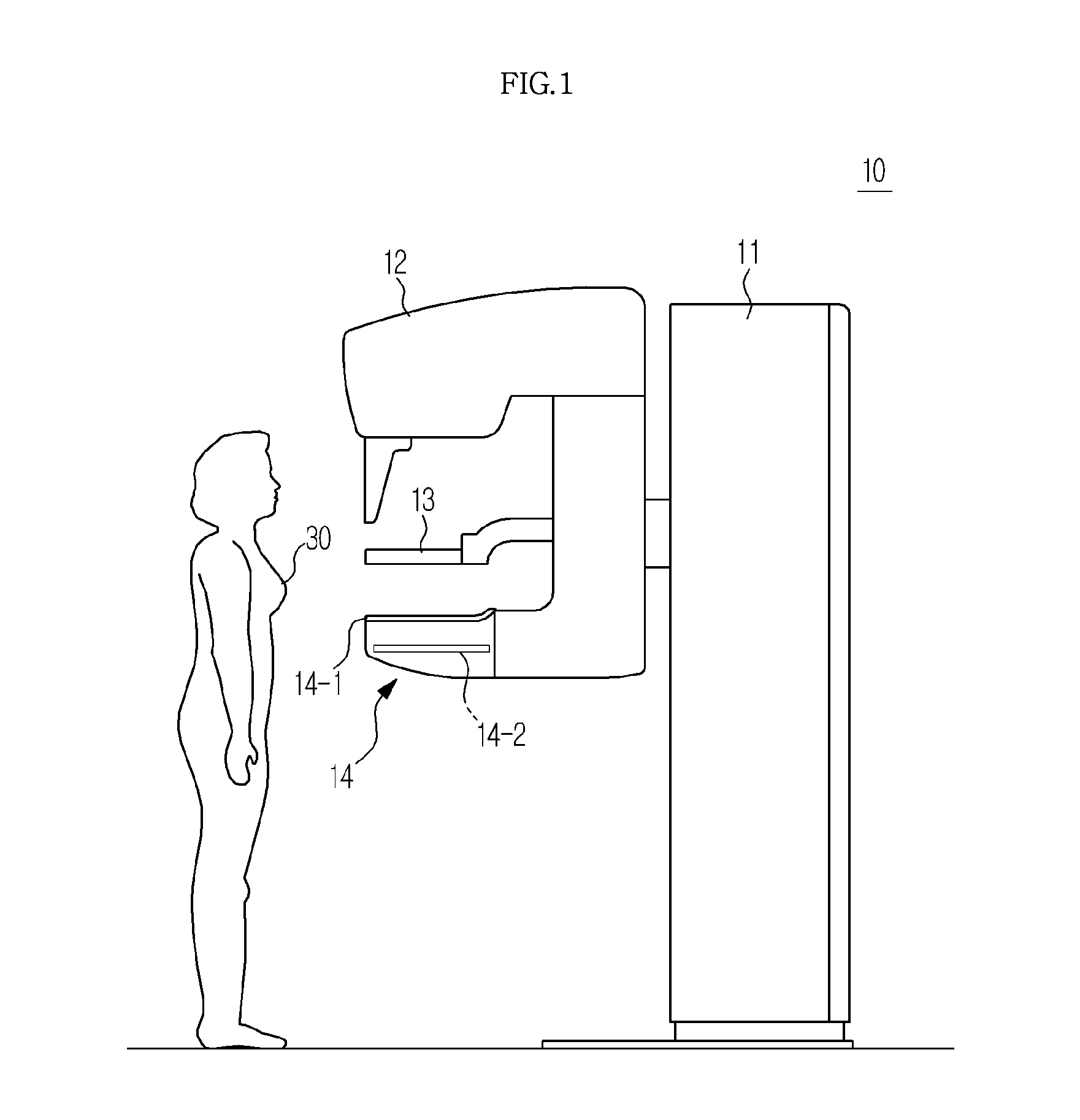
[0115]FIG. 1 is a view illustrating an external appearance of an X-ray imaging device used for breast X-ray imaging.

[0116]The X-ray imaging device 10 used for breast X-ray imaging has a structure specialized for imaging a breast, unlike a general X-ray imaging device. ...


PUM


Abstract

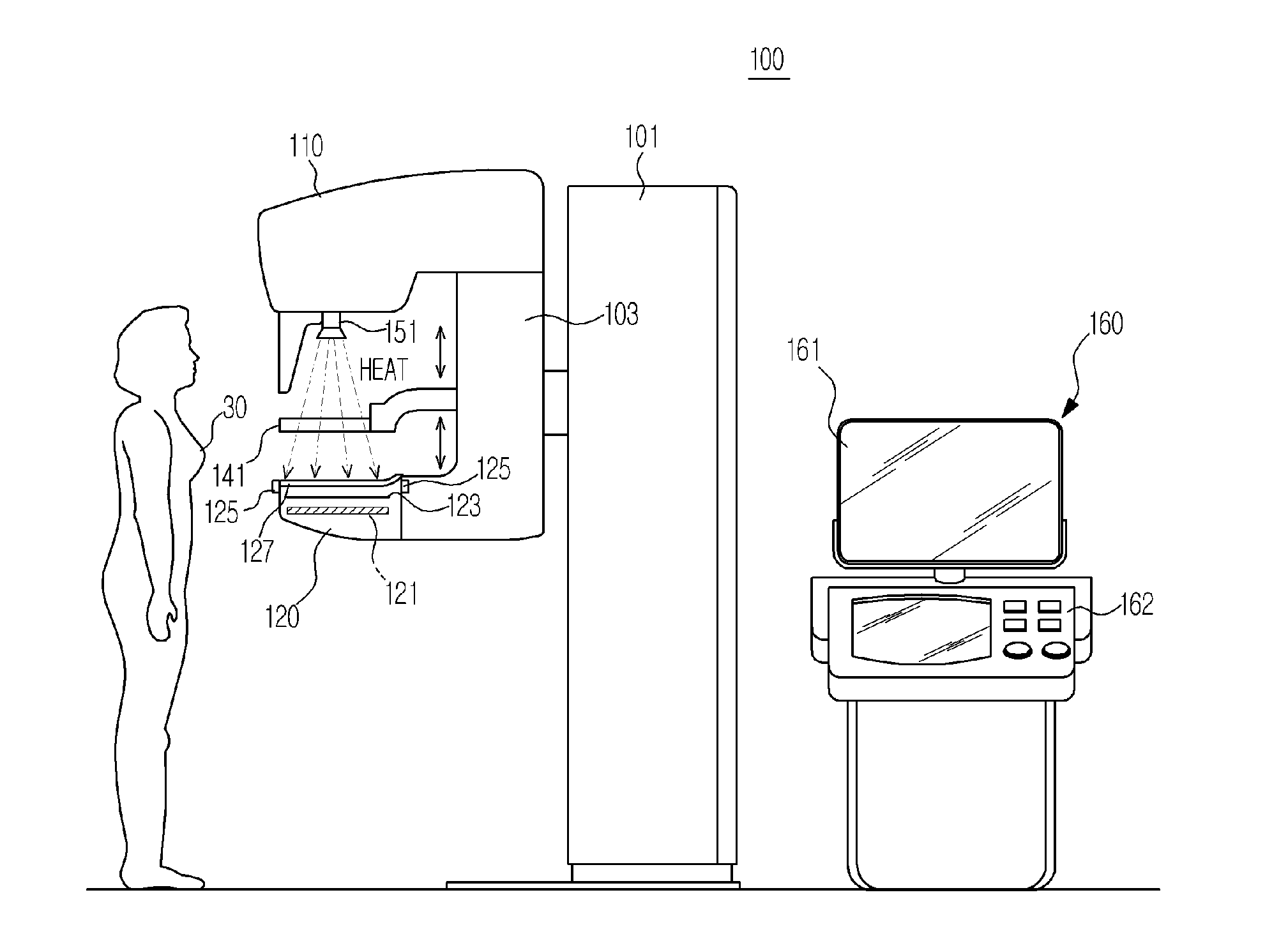
An X-ray imaging device and a method for controlling the same generates radiant heat to elevate a temperature of an area of the X-ray imaging device contacting the breast and thereby reduces patient's discomfort, and controls a temperature to maintain the temperature.The X-ray imaging device includes an X-ray source to generate X-rays and emit the X-rays to an object, an X-ray detector to detect X-rays transmitted through the object and convert the X-rays into an electrical signal, an object contact area mounted on the X-ray detector a compression paddle to compress the object disposed on the object contact area, a heat source to generate heat and transfer the heat to at least one of the object contact area and the compression paddle, and an insulation member formed between the object contact area and the X-ray detector to block transfer of heat to the X-ray detector.

Description

CROSS-REFERENCE TO RELATED APPLICATIONS[0001]This application claims the benefit of Korean Patent Application No. 10-2013-0033431 filed on Mar. 28, 2013 in the Korean Intellectual Property Office and Korean Patent Application No. 10-2014-0000901 filed on Jan. 3, 2014 in the Korean Intellectual Property Office, the disclosures of which are incorporated herein by reference.BACKGROUND[0002]1. Field[0003]One or more embodiments relate to an X-ray imaging device for forming an X-ray image by radiating X-rays to an object, in particular, a breast, and a method of controlling the same.[0004]2. Description of the Related Art[0005]An X-ray imaging device acquires an inner image of an object by radiating X-rays to the object and analyzing X-rays having transmitted through the object. Because transmittance of X-rays varies depending on a material constituting the object, an inner structure of the object is imaged by detecting a level or intensity of X-rays having transmitted through the object...

Claims


Application Information

Patent Timeline
Patent Type & Authority Applications(United States)
IPC IPC(8): A61B6/00
CPCA61B6/502A61B6/54A61B6/0414A61B6/44A61B6/08
Inventor CHOI, JIN HO
Owner SAMSUNG ELECTRONICS CO LTD
Learn More