Endodermal cells from pluripotent cells
a technology of endodermal precursor cells and pluripotent cells, applied in cell culture active agents, cell culture cells, biochemistry apparatus and processes, etc., can solve the problems of limited endodermal precursor cells, current therapies based on organ transplantation, and modest results to da
- Summary
- Abstract
- Description
- Claims
- Application Information
AI Technical Summary
Benefits of technology
Problems solved by technology
Method used
Image


Examples
example 1
Differentiation of Wild-Type (Wt) and G6PdΔ Murine Embryonic Stem Cells
[0024]Wt murine or with a deletion of the gene coding for G6PD (G6pdΔ) embryonic stem (ES) cells have been differentiated following the method described in Fico et al. (Fico A et al., Stem Cells Dev 2008). Briefly, 48 h before the induction of the differentiation, the ES cells have been plated onto pre-gelatinized plates. On day 0, the cells have been dissociated, thus obtaining a single cell suspension, which have subsequently been plated onto gelatinized plates at a density of 1.500 cells / cm2. During the differentiation process, the culture medium has been changed every other day for the first 6 days, then daily. The culture medium used consists in the medium free from integrated Knockout Serum Replacement (KSR) serum, and contains knockout Dulbecco minimal essential medium, integrated with 15% KSR (Invitrogen), 2 mM glutamine, 100 U / ml penicillin / streptomycin, and 0.1 mM β-mercaptoethanol.
[0025]From 6 days of ...
example 2
Differentiation of G6PdΔpG6pd Murine Embryonic Stem Cells
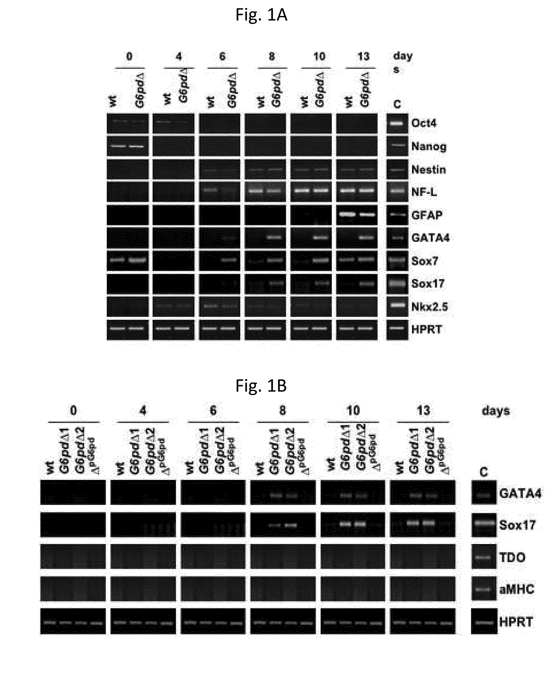
[0027]G6pdΔpG6pd cells, i.e., G6pdΔ cells transfected with an expression vector containing a puromycin-resistance gene in which the expression of the G6PD gene is under the control of the (3-actin promoter, have been used. Consequently, the G6PD expression turns out to be restored. To confirm the role played by G6PD in the endodermal differentiation, during the differentiation of G6pdΔpG6pd cells, which took place by following the same method used in Example 1, the expression of GATA4 and Sox17 has never been observed (FIG. 1B).
example 3
Induction of Defined and Extra-Embryonic Endoderm
[0028]To date, markers expressed exclusively in the definitive endoderm are not available; however, it has been shown in Borowiak et al. (supra) that the morphology acquired by Sox17-positive cells is capable of discriminating between definitive endodermal cells or extra-embryonic endodermal cells. Sox17+ cells that occur grouped belong to the definitive endoderm, while isolated Sox17+ cells belong to the extra-embryonic endoderm. In fact, the grouped Sox17 cells do not express extra-embryonic endodermal markers. During the differentiation of the G6pdΔ cells, grouped, but also dispersed, Sox17+ cells have been observed. The presence of extra-embryonic GATA4 and Sox7 markers has been confirmed (FIG. 1A) by a RT-PCR analysis, thus leading to the conclusion that during the differentiation of G6pdΔcells definitive and extra-embryonic endodermal cells are induced.
PUM
 Abstract
Description
Claims
Application Information
 - R&D
- Intellectual Property
- Life Sciences
- Materials
- Tech Scout
- Unparalleled Data Quality
- Higher Quality Content
- 60% Fewer Hallucinations
Browse by: Latest US Patents, China's latest patents, Technical Efficacy Thesaurus, Application Domain, Technology Topic, Popular Technical Reports.
© 2025 PatSnap. All rights reserved.Legal|Privacy policy|Modern Slavery Act Transparency Statement|Sitemap|About US| Contact US: help@patsnap.com