Bioscaffolds for formation of motor endplates and other specialized tissue structures
a technology of motor endplates and bioscaffolds, applied in the field of biomaterial constructs, can solve the problems of few clinical treatments available and the inability to recover muscle function, and achieve the effects of promoting muscle phenotype, promoting functional clusters, and improving function
- Summary
- Abstract
- Description
- Claims
- Application Information
AI Technical Summary
Benefits of technology
Problems solved by technology
Method used
Image
Examples
example 1
Expression of Acetylcholine Receptors
[0078]During the characterization of tissue engineered muscle repair construct (TEMR) morphology following bioreactor preconditioning, it was discovered that multinucleated cells of TEMR constructs expressed acetylcholine (ACh) receptors and, in rare cases, exhibited aggregation of these receptors.
[0079]Interestingly, the aggregation of ACh receptors in one construct is beginning to exhibit the characteristic pretzel shape of a motor endplate in mature fibers. These findings are indicative of mature TEMR constructs that produce clinically relevant force, as innervation of implanted TEMR constructs is thought to promote functional restoration of traumatically injured muscle tissue, and it is now known that these cells 1) express ACh receptors and 2) have the capacity to develop a motor endplate.
example 2
Agrin-Stimulated Acetylcholine Receptor Clustering
[0080]For proof-of-concept studies on the ability to achieve clustering of acetylcholine receptors via stimulation by agrin, a C2C12 mouse myoblast cell line was used. A 2-dimensional version of the scaffolds described above was also used to allow histological evaluation.
[0081]C2C12 cells were seeded on 2-dimensional fibrin hydrogels for 4-10 days. Separately, agrin was coated onto or covalently immobilized on the surface of polystyrene beads. At 3-6 days of C2C12 cell culture, the agrin-coated beads were placed onto the cells for 24 hours or more. Cells were then stained for alpha-bungarotoxin, a marker of acetylcholine receptor clustering.
[0082]Agrin-coated beads on C2C12 cells cultured on fibrin hydrogels resulted in areas of clustering (FIG. 2). All areas of clustering occurred where beads were present, indicating that the agrin-coated beads promoted clustering behavior. Physical adsorption of agrin onto beads was shown to be sup...
example 3
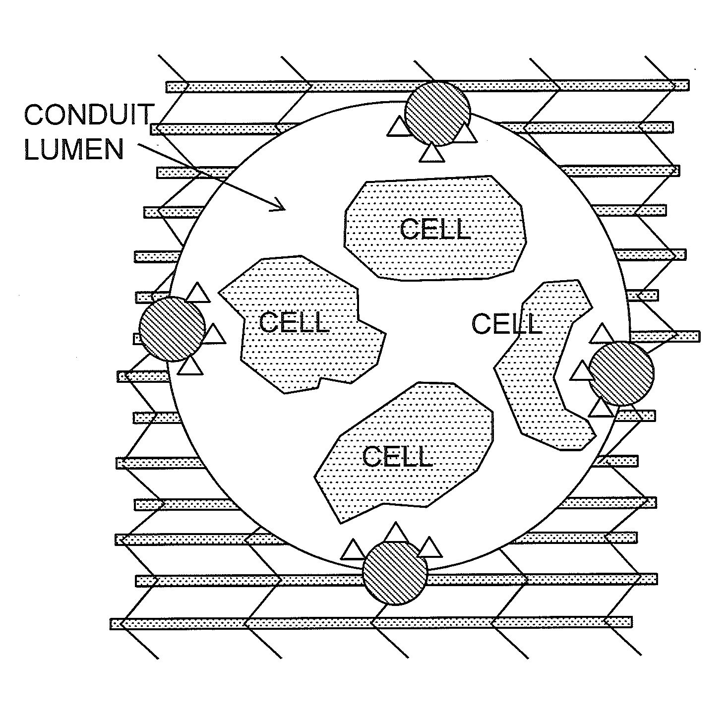
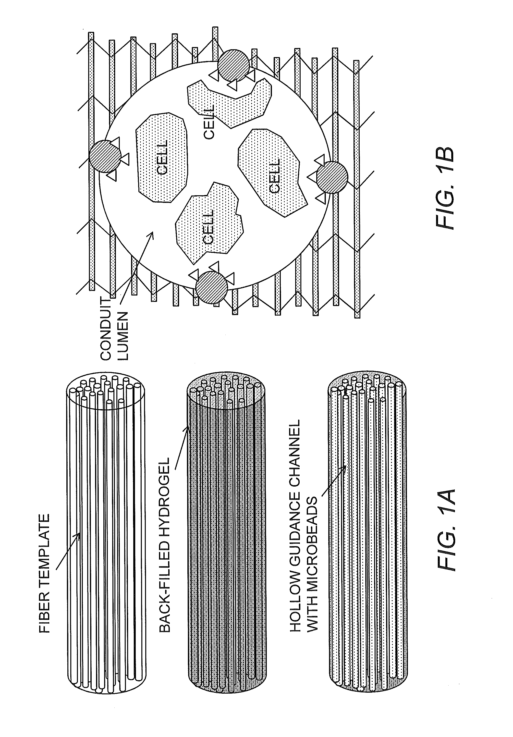
Incorporation of Agrin-Coated Beads Into Scaffolds
[0085]Agrin-coated beads are incorporated into 2-D hydrogels and the 3-D scaffolds described above (fabricated using a poly(tetrafluoroethylene) tubular or rectangular mold), myoblasts are seeded onto the formed 3-D scaffolds, and studies similar to those described above in Example 2 are conducted to determine the efficacy of cell seeding in three dimensions as well as the level of acetylcholine receptor clustering achieved when beads are part of the scaffold material. A rectangular, 3-D, patterned fibrin scaffold was seeded by static addition of a cell suspension and subsequent infiltration. As shown in FIG. 5, C2C12 cells substantially infiltrated the full thickness of the porous architecture via native cell motility, demonstrating the suitability of both the fibrin biomaterial and the sacrificial patterning process to myoblast-like cell seeding.
[0086]Furthermore, microscopy revealed that the seeded myotube-like cells fused within ...
PUM
| Property | Measurement | Unit |
|---|---|---|
| thick | aaaaa | aaaaa |
| thick | aaaaa | aaaaa |
| thick | aaaaa | aaaaa |
Abstract
Description
Claims
Application Information
 - R&D
- Intellectual Property
- Life Sciences
- Materials
- Tech Scout
- Unparalleled Data Quality
- Higher Quality Content
- 60% Fewer Hallucinations
Browse by: Latest US Patents, China's latest patents, Technical Efficacy Thesaurus, Application Domain, Technology Topic, Popular Technical Reports.
© 2025 PatSnap. All rights reserved.Legal|Privacy policy|Modern Slavery Act Transparency Statement|Sitemap|About US| Contact US: help@patsnap.com


