Method for proliferation cardiomyocytes using micro-rna

a cardiomyocyte and micro-rna technology, applied in the field of proliferating cardiomyocytes using micro-rnas, can solve the problems of further exhaustion and death of cardiomyocytes, poor prognosis of patients with heart disease, and consequent impairment of myocardial function, and achieve the effect of marked proliferation activity

Inactive Publication Date: 2014-07-31
DAIICHI SANKYO CO LTD
View PDF1 Cites 0 Cited by
  • Summary
  • Abstract
  • Description
  • Claims
  • Application Information

AI Technical Summary

Benefits of technology

[0083]By using the miRNAs according to the present invention, one can provide a method for proliferating cardiomyocytes, a vector for use in the treatment of heart disease, and a pharmaceutical composition for treating heart disease. When the method of proliferating cardiomyocytes according to the present invention is used, cell division of cardiomyocytes can be induced for cell proliferation to take place more efficiently than in the prior art, and the cardiomyocyes prepared by this method can be utilized as cells for the screening of various drugs and for transplantation therapy. In addition, by applying this method in gene therapy, heart diseases due to necrosis, depletion or other failure of cardiomyocytes may potentially be treated in an attempt as part of regenerative therapy. The miRNAs according to the present invention have been shown to exhibit a marked proliferation activity on cardiomyocytes.

Problems solved by technology

In consequence, the remaining cardiomyocytes try to maintain the cardiac function through compensatory hypertrophy, but if this condition continues and exceeds tolerable threshold of the myocardial tissue, it leads to further exhaustion and death of cardiomyocytes and a consequent impairments myocardial function, namely, heart failure.
In addition, patients with heart disease have a very poor prognosis and their five year survival rate is only about 50%.
Conventionally used therapeutics for heart failure are cardiotonic drugs such as digitalis and xanthine preparations that increase the force of myocardial contraction but these drugs are known to potentially worsen rather than ameliorate the diseased condition if administered over a prolonged period of time.
In recent years, treatments with drugs such as β-blockers and ACE inhibitors that reduce an excessive cardiac load due to elevations of the sympathetic nervous system or rennin-angiotensin system are becoming the mainstream but these methods are just symptomatic and unable to restore the damaged cardiac tissue itself.
Although heart transplantation is a radical therapy for severe heart failure, it involves various problems such as the scarcity of donor organs, sensitive medical ethics, and the high costs of physical or economic burdens on patients, making it difficult to take this approach as a common therapy.

Method used


Image

  • Method for proliferation cardiomyocytes using micro-rna
  • Method for proliferation cardiomyocytes using micro-rna
  • Method for proliferation cardiomyocytes using micro-rna

Examples


example 1

BrdU-Uptake-Based Screening of miRNA Library Using Primary Culture of Cardiomyocytes

[0153]The heart was excised from 2-4 day old rats (Wistar rats obtained from SHIMIZU Laboratory Supplies Co., Ltd.) and treated with collagenase to prepare a cell population, from which cardiomyocyte fractions were recovered by Percoll density gradient centrifugation (a Percoll suspension of cells with a density of 1.082 g / mL was layered on a Percoll solution with a density of 1.050 g / mL or 1.060 g / mL, and subjected to centrifugation at 4° C. and 3000 rpm (2000 G) for 25 minutes). The thus obtained cardiomyocytes were suspended in an Eagle minimum medium (nakalai tesque) supplemented with 5% fetal calf serum (FCS; SAFC Bioscience) and then seeded on culture dishes for cell culture in a CO2 incubator at 37° C. The thus prepared cardiomyocytes (15,000 cells / well on a 96-well plate) were transfected with 2 pmol each of Pre-miR™ miRNA Precursor Library-HumanV2 (Ambion: a library designed to mimic the 328...

example 2

Analysis of a Marker for the Progress of Cell Cycle by Forced miRNA Expression in Primary Culture of Cardiomyocytes

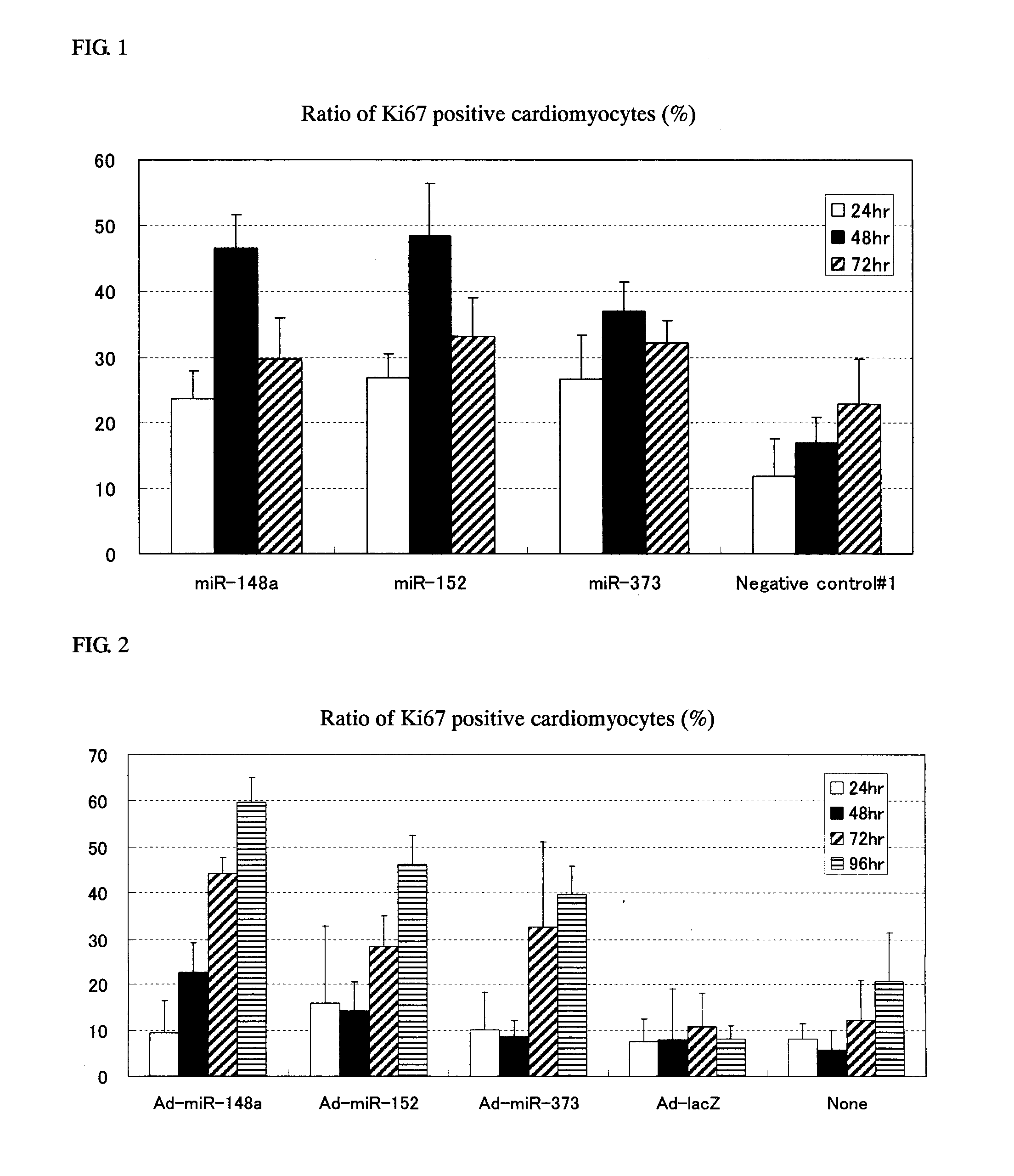
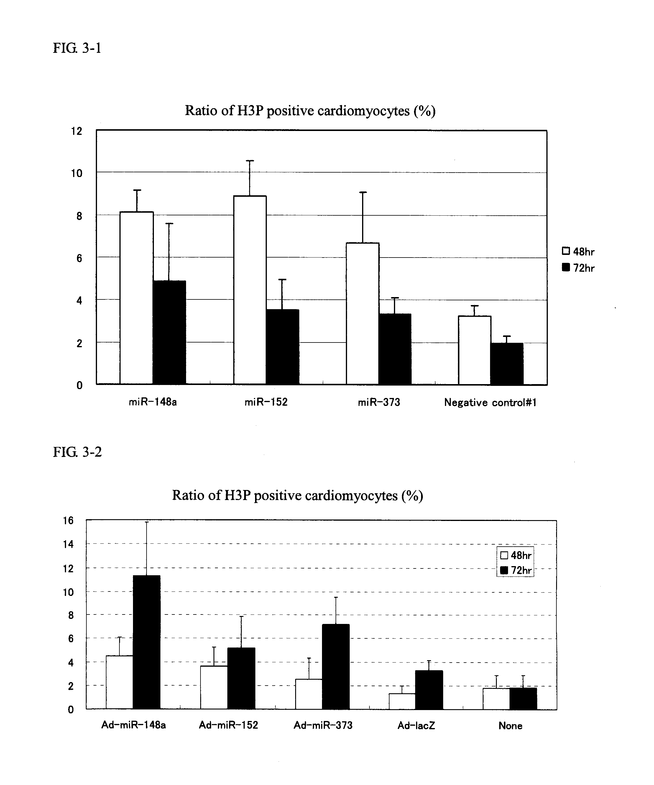
[0154]Cardiomyocytes (100,000 cells / well on a 24-well plate) were transfected with 1 pmol of Pre-miR miRNA Precursor Molecules (Ambion) that were designed to mimic miR-148a, miR-148b, miR-152 and miR-373 (the candidate mature miRNAs obtained in Example 1) and 1 pmol of Negative control #1 (a negative control, also available from Ambion) using Lipofectamine™ RNAiMAX (Invitrogen) and 2-4 days after the transfection, the cells were fixed with 4% paraformaldehyde.

[0155]Following subsequent reaction with anti-Ki67 antibody (Thermo SCIENTIFIC) (1:200 dilution) and anti-Troponin T antibody (Thermo SCIENTIFIC) (1:200 dilution), the cells were stained with Alexa Fluor™ labeled antibody (Alexa-488 or Alexa-568; Molecular Probes) (both 1:400 dilutions). The cell nucleus was stained with a 4,6-diamidine-2′-phenylindole diydrochloride: DAPI) solution (1 μg / mL).

[0156]The cardiomyocyt...

example 3

Analysis of a Marker for the Progress of Cell Cycle by Adenovirus-Forced miRNA Expression in Primary Culture of Cardiomyocytes

[0159]Adenoviral vector for expressing miR-148a, miR-152, and miR-373 were prepared by using ViraPower™ Adenoviral Expression System (Invitrogen). To be more specific, human genomic DNA fragments comprising the precursor sequences of the respective miRNAs and adjacent several hundred nucleotides were amplified by PCR using the primers shown below, with the template being human genomic DNAs.

hsa-miR-148a-F:(SEQ ID NO: 11)5′-caccgaacacacctgcaggaagaaact-3′hsa-miR-148a-R:(SEQ ID NO: 12)5′-gttcccatttacagggtttaaccca-3′hsa-miR-152-F:(SEQ ID NO: 13)5′-caccgtcccagactcggctcccatca-3′hsa-miR-152-R:(SEQ ID NO: 14)5′-actcgaggtggacaccctgtgt-3′hsa-miR-373-F:(SEQ ID NO: 15)5′-caccgtgaccaaggggctgtatgca-3′hsa-miR-373-R:(SEQ ID NO: 16)5′-ctgcccaccccagaatatgcca-3′.

[0160]The resulting PCR products were inserted into a pENTR / D-TOPO vector (Invitrogen) and after checking their base s...


PUM

PropertyMeasurementUnit
densityaaaaaaaaaa
densityaaaaaaaaaa
densityaaaaaaaaaa

Abstract

The present invention addresses the problems of providing a method for proliferating cardiomyocytes using a miRNA that promotes the proliferation of cardiomyocytes, a vector for use in the treatment of heart disease, a pharmaceutical composition for treating heart disease, and so forth.The present invention provides a method for proliferating cardiomyocytes using a miRNA having the cardiomyocyte proliferation promoting action, a vector for use in the treatment of heart disease that comprises said miRNA, a pharmaceutical composition for treating heart disease that comprises said vector, and so forth. A particularly preferred miRNA is one that is selected from the group consisting of mature miRNAs, i.e., miR-148a, miR-148b, miR-152, and miR-373, and precursors of said miRNAs, as well as variants and analogs thereof.

Description

TECHNICAL FIELD[0001]The present invention relates to a method for proliferating cardiomyocytes using microRNAs, a vector for treating heart disease, and a pharmaceutical composition for treating heart disease.BACKGROUND ART[0002]In adults, cardiomyocytes have lost their ability to divide, so once they become necrotic or lost entirely upon exposure to various stresses such as ischemia or myocarditis, the lost cardiomyocytes can be never regenerated. In consequence, the remaining cardiomyocytes try to maintain the cardiac function through compensatory hypertrophy, but if this condition continues and exceeds tolerable threshold of the myocardial tissue, it leads to further exhaustion and death of cardiomyocytes and a consequent impairments myocardial function, namely, heart failure.[0003]Heart diseases including heart failure are the second most common cause of death among Japanese people. In addition, patients with heart disease have a very poor prognosis and their five year survival...

Claims


Application Information

Patent Timeline
Patent Type & Authority Applications(United States)
IPC IPC(8): C12N15/113C12N5/077A61K35/34
CPCA61K31/7105A61K31/7115A61K31/712A61K31/7125C12N5/0657C12N15/113C12N2310/141C12N2320/11A61K35/34A61P9/00A61P9/04A61P9/10A61K48/00
Inventor KAWASHIMI, KAYOKOKOSHIMIZU, UICHI
Owner DAIICHI SANKYO CO LTD
Why Eureka
Try Eureka