Apparatus and methods for photosensor quadrant sharing

a quadrant and photosensor technology, applied in the field of apparatus and methods for photosensor quadrant sharing, can solve the problems of poor practical clinical resolution, untapped cancer-application potential of pet in whole body, and current higher cost of sensors than pmt, so as to achieve higher resolution pqs, less cost, and economic

Active Publication Date: 2014-05-22
BOARD OF RGT THE UNIV OF TEXAS SYST
View PDF5 Cites 27 Cited by
  • Summary
  • Abstract
  • Description
  • Claims
  • Application Information

AI Technical Summary

Benefits of technology

[0025]As explained in more detail below, exemplary embodiments of the present disclosure enable the lower cost, higher resolution PQS detector design to be used to make PET detector rings and polygonal SPECT detector panels more economically and with no detection gaps. Such a configuration can provide increasing detection sensitivity, and improving data sampling and reliability without expensive, high-precision camera rotation movement. Exemplary embodiments of the present disclosure can utilize the PQS configuration illustrated in FIGS. 3 and 4.
[0026]Embodiments of the current disclosure also reduce the number of photosensors used in the camera from the original panelized PQS design. As explained in further detail below, the original PQS design would waste one half of the photosensors along each edge of each panel. In exemplary embodiments of this disclosure with a gapless continuous ring, such waste is eliminated.
[0027]Unlike existing embodiments described in the previous section, in the PQS design the light signal is confined to go to just four PMT, thus having lower pileup of event signals. Accordingly, there are several advantages exemplary embodiments in forming a gapless PQS PET detector ring compared with other existing systems that incorporate a ring that is nearly gapless.

Problems solved by technology

Despite this success, the cancer-application potential of PET in whole body is still largely untapped today because of high scanner cost, and low image resolution.
As of 2009, clinical PET cameras have imaging resolution of 4.0-6.3 mm, but because of low sensitivity, the practical clinical resolution is worse (7-10 mm), which can miss smaller (early) primary lesions and metastases.
Although other solid-state or semiconductor photosensors are also being investigated, such sensors are currently more expensive than PMT.
PMT cost is a major component cost in a PET detector system.
However, since there are only a limited effective surface finishes that can be used, the number of crystals that can be decoded by the four PMT is more limited.
The smaller number of crystal pixels in a block / array leads to lower imaging resolution.
The detection gaps are not desirable for several reasons: (a) lower detection sensitivity, (b) inadequate data sampling unless the detection system rotates, which increases mechanical complexity, reliability, positioning errors and production cost.
For the new “time-of-flight” PET applications, the detection gap without rotating the detection system is especially problematic in the image reconstruction process.
While the configuration shown in FIG. 7 addresses some of the shortcomings of prior systems, it also results in high manufacturing costs for large scale systems.
However, there is degradation in image resolution even for the region near the center of the field-of-view and sensitivity loss caused by the gaps between two detector panels.
While an accurate rotation gantry can be used to rotate the apparatus and fill the gaps of missing LOR in the sinograms, such an implementation also increases the production cost.
In such embodiments, the light guide (a) absorbs light signal and (b) reduces the light going to the PMT by a wide area light dispersion (larger than the seven PMT used for extracting the scintillating position), thereby reducing the positioning signal strength and positioning accuracy.
Furthermore, the thick light guide utilized in other embodiments absorbs and loses more light compared to gapless PQS designs that do not use an external light guide.
But for a human-scale PET system, with a hundred times more detector blocks, such individual block grinding can be prohibitively expensive.

Method used


Image

  • Apparatus and methods for photosensor quadrant sharing
  • Apparatus and methods for photosensor quadrant sharing
  • Apparatus and methods for photosensor quadrant sharing

Examples


Embodiment Construction

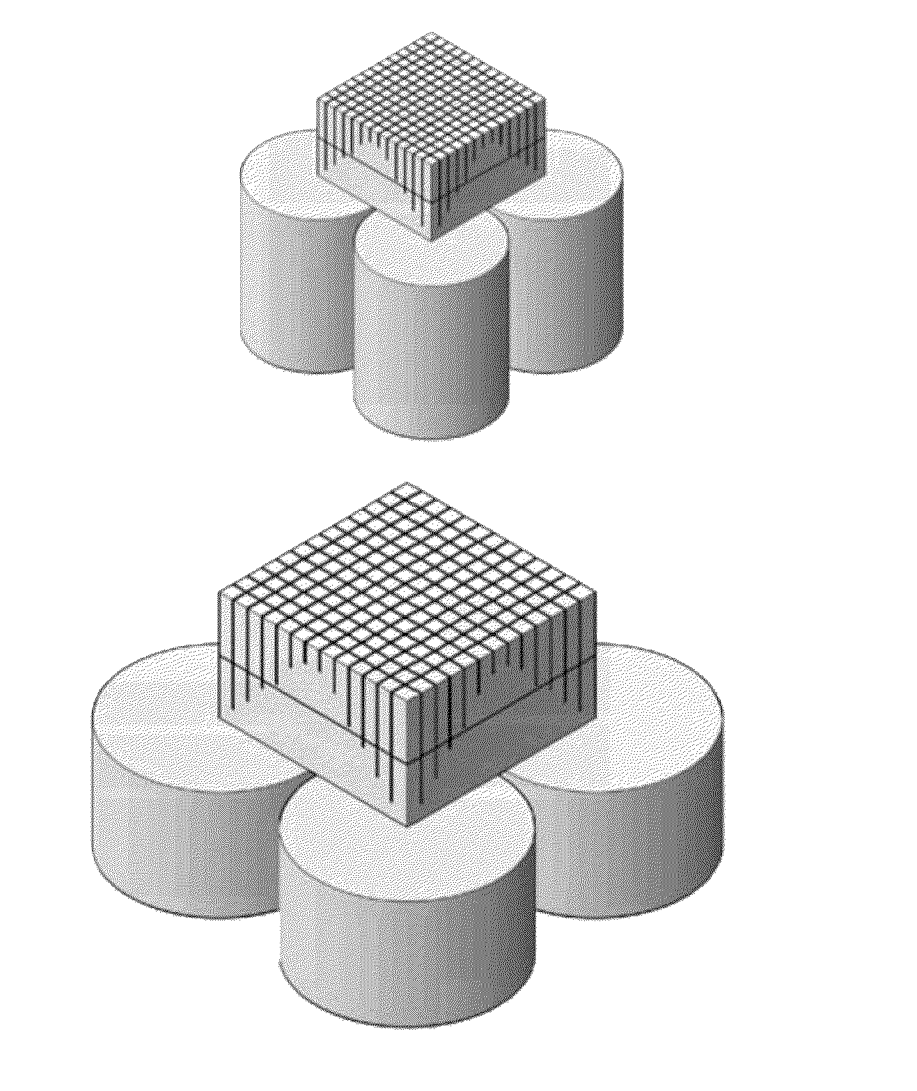
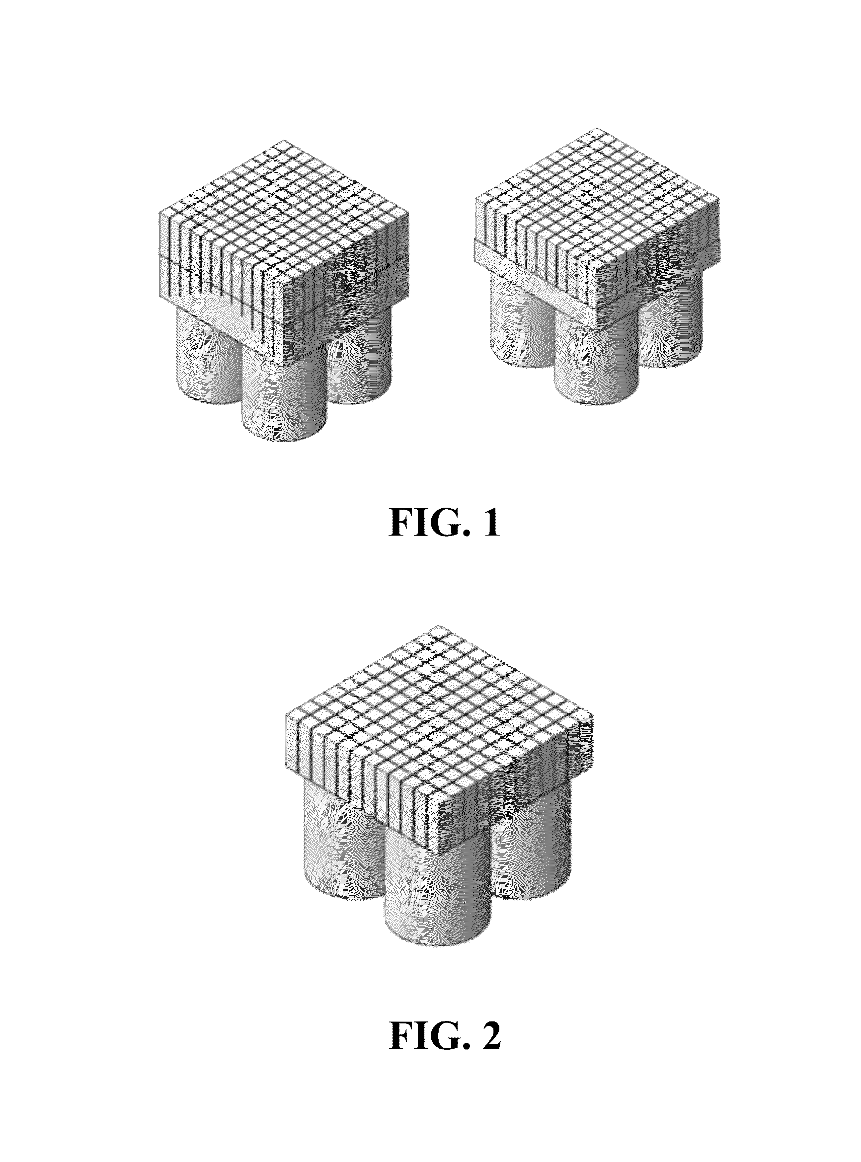
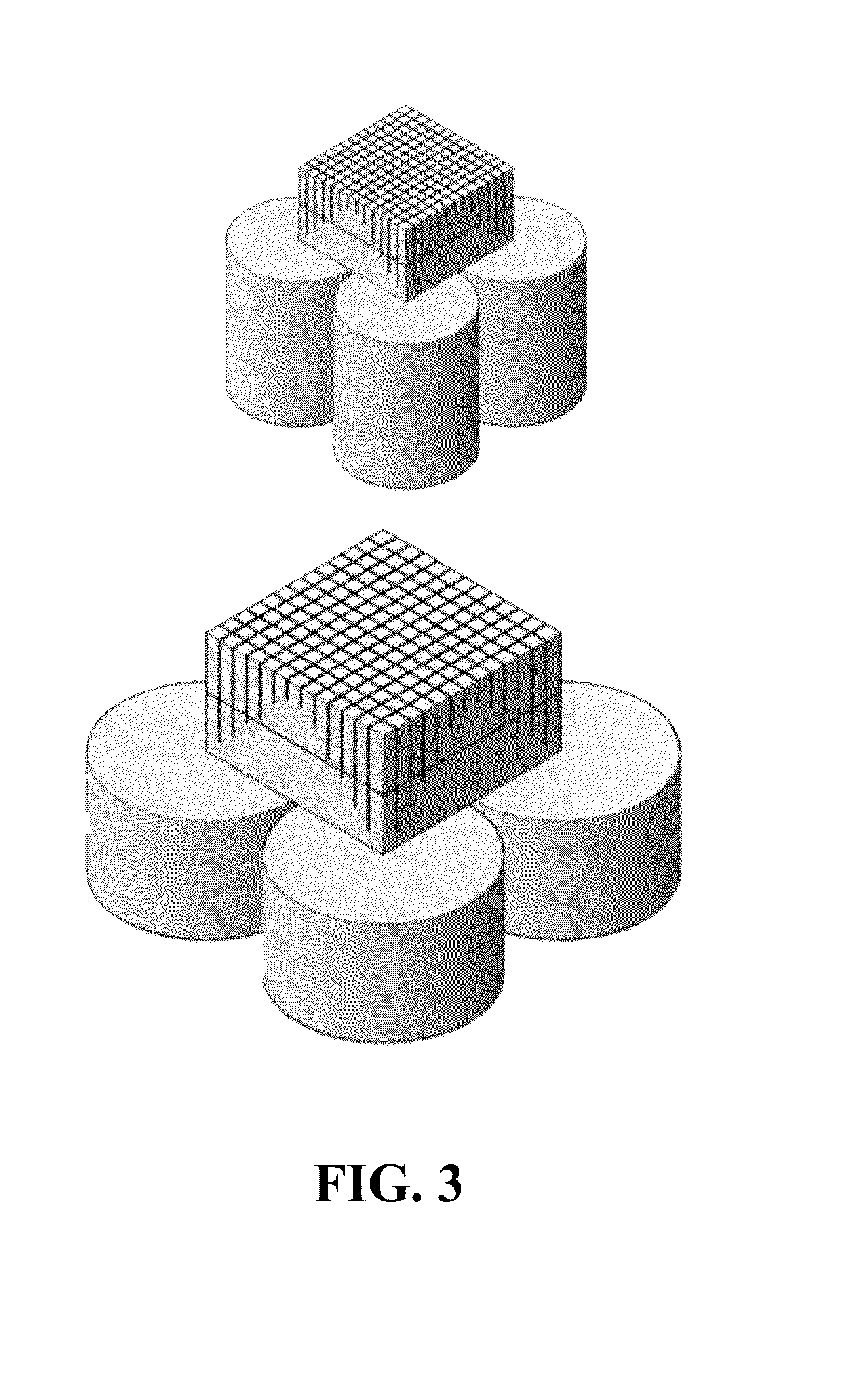
[0067]Referring now to FIGS. 9-11, a plurality of rectangular cross-section scintillation crystal blocks 510, 520, 530 and 540 are used to form an array 550 of blocks, as described in further detail below. In this exemplary embodiment, blocks 510 and 540 will be modified to have an asymmetric pentagonal cross-section, while blocks 520 and 530 will maintain their rectangular cross-section. Referring specifically now to FIG. 9, block 510 comprises a first side 511, a second side 512, a third side 513 and a fourth side 514. Similarly, block 540 comprises a first side 541, a second side 542, a third side 543 and a fourth side 544. In this embodiment, blocks 510 and 540 can be modified by tapering sides 513 and 514 and sides 543 and 544, respectively. In a specific embodiment, sides 513, 514 and 543, 544 can be tapered by grinding the sides. For purposes of clarity, the modification of block 510 will be described in detail below. It is understood that in this embodiment, the modification...


PUM


Abstract

Apparatus and methods comprising a positron emission tomography detector having a first rectangular cross-section crystal block positioned between a first pentagonal cross-section crystal block and a second pentagonal cross-section crystal block.

Description

[0001]The present application claims benefit of priority to U.S. Provisional Application Ser. No. 61 / 728,327, filed Nov. 20, 2012, the entire contents of which are hereby incorporated by reference.[0002]This invention was made with government support under contracts 5RO100EB4840 and 2RO1EB001038 awarded by NIH / NIBIB. The government has certain rights in this invention.BACKGROUND INFORMATION[0003]Positron emission tomography (PET) can detect tumor in vivo based on the living chemistry of cancer tissues. In recent years, PET has well demonstrated its broad clinical utilities in cancer diagnosis and is recognized as an important tool to study cancer functions in vivo, because of its unique ability to elicit molecular functions. The in vivo molecular imaging ability of PET has triggered considerable cancer-research interest and radiotracer development to study cancer related molecular processes such as angiogenesis, apoptosis, cell proliferation, hypoxia, gene expression and blood flow....

Claims


Application Information

Patent Timeline
Patent Type & Authority Applications(United States)
IPC IPC(8): G01T1/20
CPCG01T1/2006G01T1/1644A61B6/037
Inventor LI, HONGDIWONG, WAI-HOIZHANG, YUXUAN
Owner BOARD OF RGT THE UNIV OF TEXAS SYST
Why Eureka
Try Eureka