
Induced malignant stem cells

a stem cell and malignant technology, applied in the field of induced malignant stem cells, can solve the problems of abnormal gene expression, difficult or even impossible to determine, and difficult to retain aberrations, and achieve the effect of high or low degree of methylation

Inactive Publication Date: 2014-05-15
ISHIKAWA
View PDF6 Cites 77 Cited by
  • Summary
  • Abstract
  • Description
  • Claims
  • Application Information

AI Technical Summary

Benefits of technology

The present invention provides an induced malignant stem cell that can be cultured in the laboratory and has specific genetic or epigenetic abnormalities associated with cancer, such as methylation, mutations, or abnormal expression of genes, noncoding RNA, or proteins related to cancer. The stem cell also has abnormal metabolism or sugar chain, or instability of microsatellites in its genomic DNA. This stem cell can be used to develop new treatments for cancer and related diseases.

Problems solved by technology

However, even in the absence of such gene transduction, the cancer cell lines established in common conventional media significantly generate in vitro artifact aberrations during extended culture, including chromosomal aberrations (e.g. dislocation and deletion), genomic aberrations (genetic mutations), and epigenetic aberrations which might lead to abnormal gene expression (Non-Patent Document 10).
This gives rise to a problem that it is difficult to retain the aberrations such as mutations that occurred in cancer cells which were inherent causes of carcinogenesis or malignant transformation in vivo as such within the cells while minimizing the in vitro artifact aberrations.
In cancer therapy research and the research for cancer-related drug discovery, even if the genomic or epigenetic aberrations in the cancer cell lines established by extended culture in such conventional media are analyzed, it is extremely difficult or even impossible to determine whether those aberrations were inherent in mammalian cancer cells as an etiology of carcinogenesis or malignant transformation, or in vitro artifact aberrations that occurred during culture and, hence, it is difficult to unravel an appropriate etiology of carcinogenesis or malignant transformation on the basis of the results of those analyses.
It has been inappropriate to use such cells to search for a target in the discovery of a cancer therapeutic drug, screen for a candidate for cancer therapeutic drug, and the like.
A further problem is that despite the fact that cancer stem cells are highlighted as an important target in drug discovery, the cancer cells that are contained in a fresh cancer tissue make up a hierarchical and heterogeneous cell population and it is not easy to identify which cancer cells are cancer stem cells.
Recently, there was reported a study for identifying cancer stem cells from a cancer cell line or primary cultured cancer cells (Non-Patent Document 11) but there is no report of successful in vitro proliferation and extended culture of monoclonal cancer cells, nor has been reported any technology by which they can be proliferated and subjected to in vitro expansion culture until their number reaches the necessary level for application in drug discovery and for use in cancer research.
However, when a heterogeneous cancer tissue which is hierarchical and is not clonal is analyzed, data for the average genome of the cancer cell populations involved or the genome of the most abundant cancer cell population will be presented as a result but the problem is that it cannot be positively determined whether the result originates from the cancer cells in the cancer tissue that are an etiology of malignant transformations (development and metastasis).
However, as of today, no cells have been established that correspond to the results of analyses and which are amenable to expansion culture and it has been impossible to perform functional analysis, XENOGRAFT modeling, and target / compound screening in drug discovery using the available cell lines.

Method used


Image

  • Induced malignant stem cells
  • Induced malignant stem cells
  • Induced malignant stem cells

Examples


example 1

Preparation of Induced Malignant Stem Cells from Cells (GC2) Derived from Cancer Tissues of a Gastric Cancer Patient

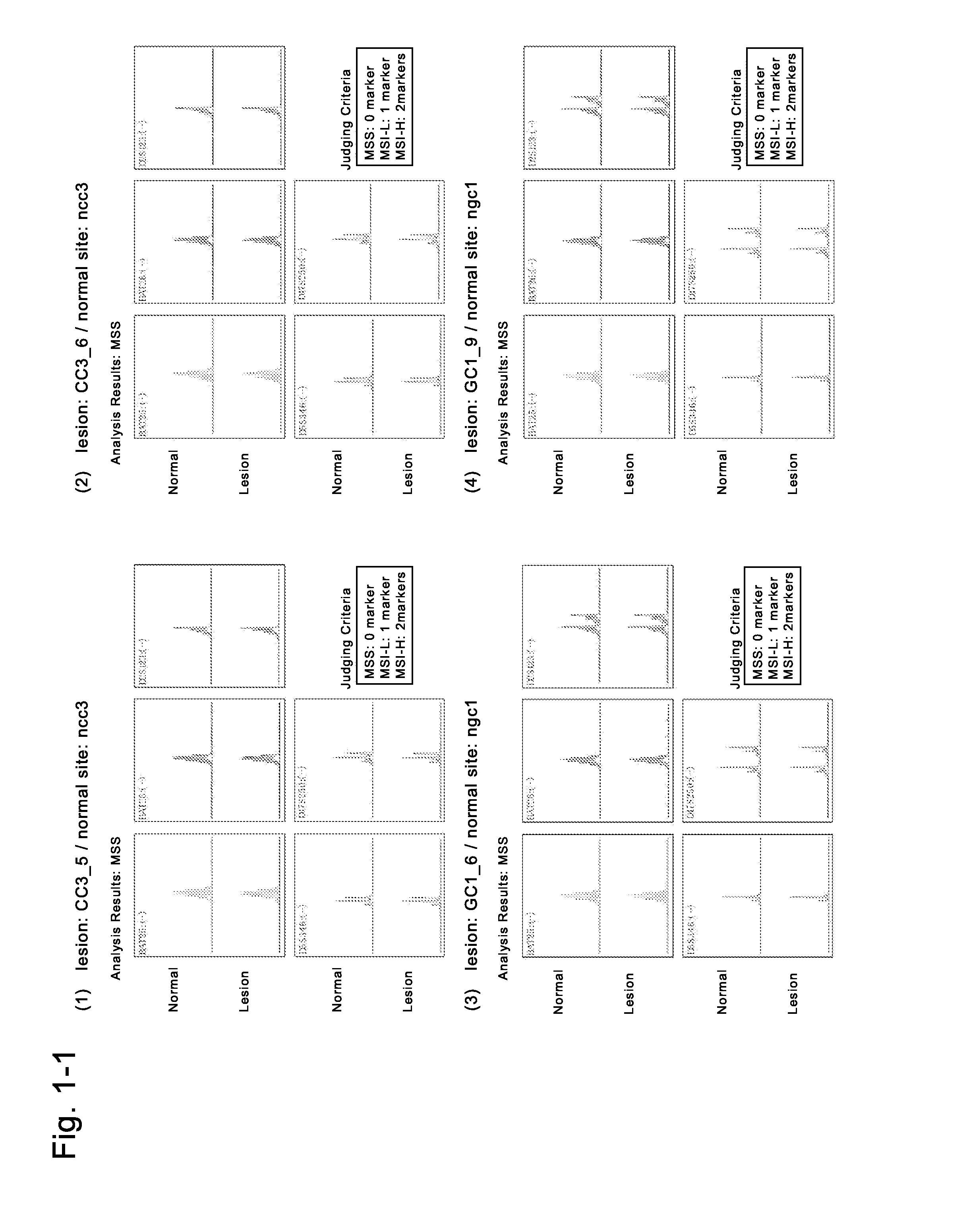
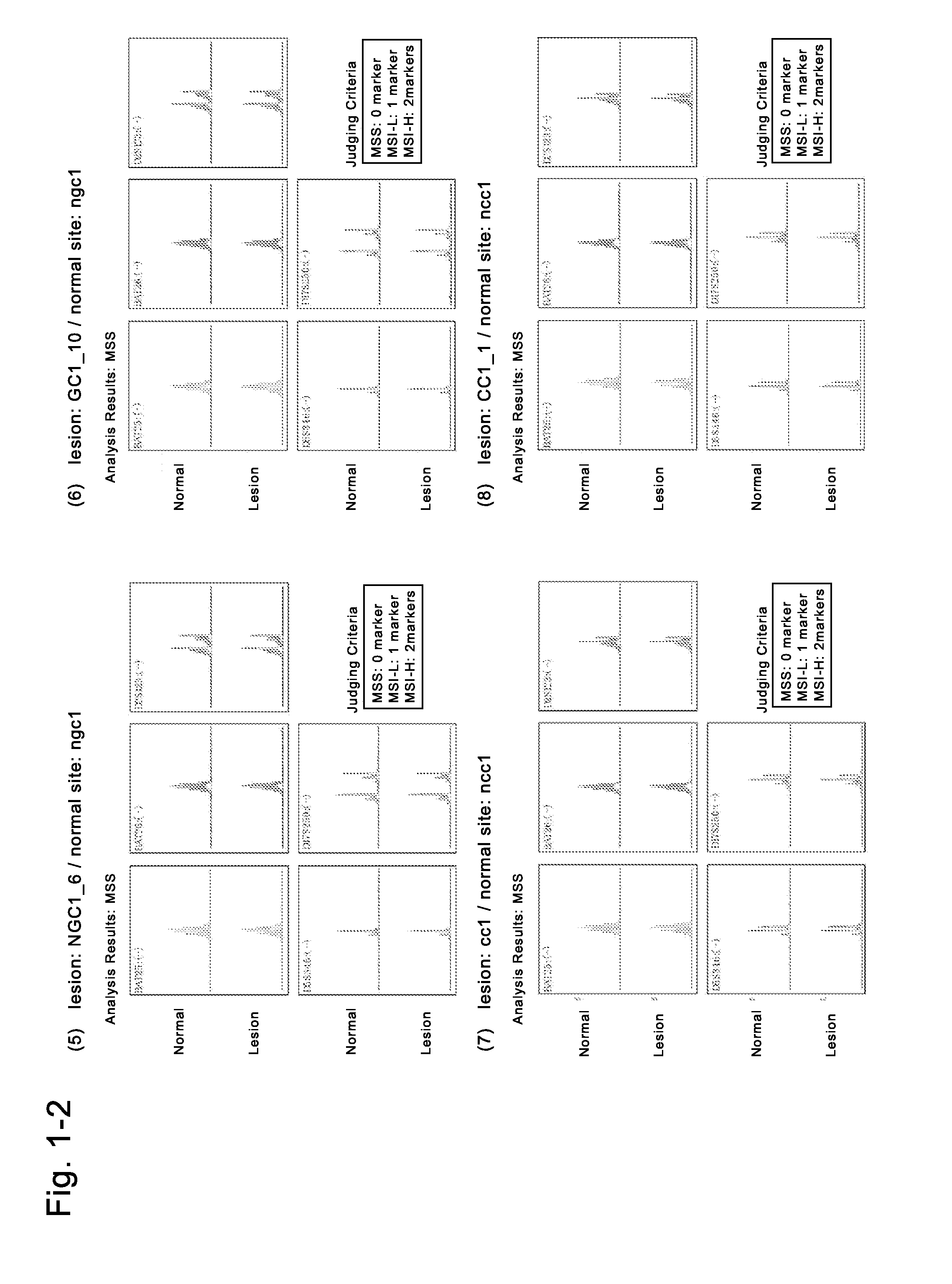
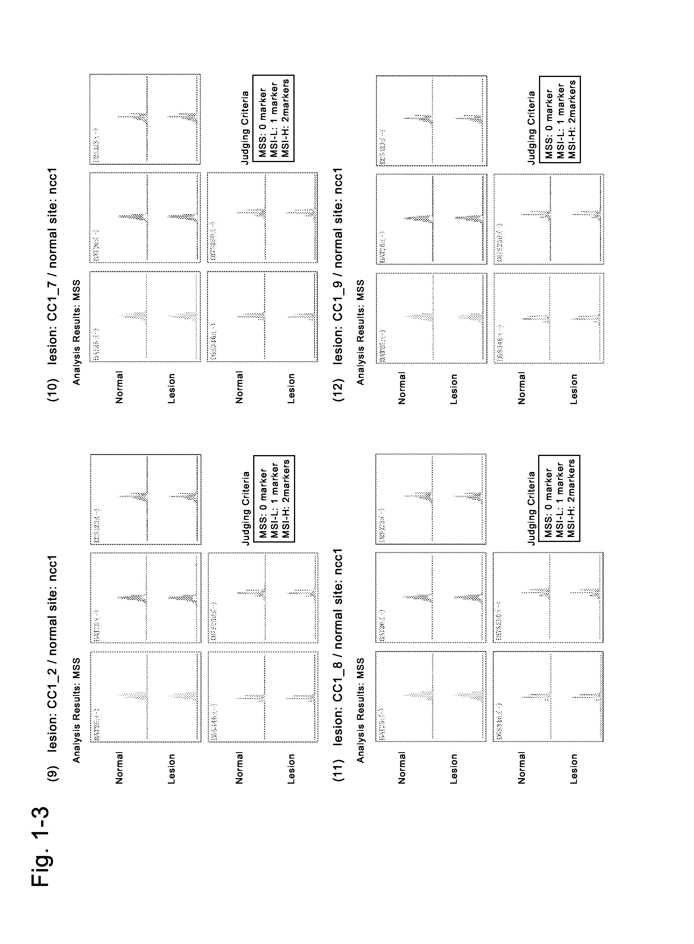
[0220]The fresh cancer tissues of a gastric cancer patient of donor No. 1 (medical information: a 67-year-old Japanese woman with a gastric cancer, blood type O, no chemotherapy, no radiotherapy, no immunosuppressive therapy, no smoking history, no drinking history, no drug addiction, no drug therapy, HIV-negative, HCV-negative, HBV-negative, syphilis-negative) which had been refrigerated for several hours and transported in a preservation solution (Hanks' solution supplemented with kanamycin and Fungizone) were used to isolate cells (GC2). The fresh non-cancer tissues of the patient were also used to isolate cells (NGC2). To the resultant cells derived from the gastric (solid) cancer tissues, the solution of the four Sendai viral vectors containing any of four genes (POU5F1, KLF4, SOX2, c-Myc) (DNAVEC CytoTune iPS (DV-0301-1)) was added for genetic transduction, where...

example 2

Preparation of Human Induced Malignant Stem Cells from Cells (CC3) Derived from Cancer Tissues of a Colon Cancer Patient

[0234]The fresh cancer tissues of a colon cancer patient of donor No. 2 (medical information: a 77-year-old Japanese man with a sigmoidal colon cancer, blood type A, no chemotherapy, no radiotherapy, no immunosuppressive therapy, no smoking history, drinking history: 1 bottle of beer / day, no drug addiction, no drug therapy, HIV-negative, HCV-negative, HBV-negative, syphilis-negative) which had been refrigerated for several hours and transported in a preservation solution (Hanks' solution supplemented with kanamycin and Fungizone) were used to isolate cells (CC3). The fresh non-cancer tissues of the same donor were also used to isolate cells (NCC3). To the resultant cells derived from the cancer tissues of the colon cancer patient, the solution of the four Sendai viral vectors containing any of four genes (POU5F1, KLF4, SOX2, c-Myc) (DNAVEC CytoTune iPS (DV-0301-1))...

example 3

Preparation of Retroviral Vectors

[0244]The plasmids of the three retroviral vectors containing any of three genes, POU5F1-pMXs, KLF4-pMXs, and SOX2-pMXs, were transduced into Plat-GP cells (packaging cells for preparing a pantropic retroviral vectors) using Fugene HD (Roche; Cat No. 4709691) to thereby prepare solutions of the retroviral vectors. The details of the procedure are as described below.

[0245]

[0246]POU5F1-pMXs, KLF4-pMXs, and SOX2-pMXs were the constructed vectors (Table 9).

[0247]The amounts of the respective vectors were as follows: 5 μg of POU5F1-pMXs, 2.5 μg of KLF4-pMXs, 1.25 μg of SOX2-pMXs, 1.25 μg of Venus-pCS2, 5 μg of VSV-G-pCMV, 1.25 μg of GFP-pMXs (Cell Biolab), and 45 μL of FuGENE HD.

[0248]

[0249]POU5F1-pMXs, KLF4-pMXs, and SOX2-pMXs were the constructed vectors (Table 9).

[0250]The amounts of the respective vectors were as follows: 5 μg of POU5F1-pMXs, 2.5 μg of KLF4-pMXs, 1.25 μg of SOX2-pMXs, 1.25 μg of Venus-pCS2, 5 μg of VSV-G-pCMV, 1.25 μg of GFP-pMXs, and...


PUM

PropertyMeasurementUnit
Fractionaaaaaaaaaa
Fractionaaaaaaaaaa
Timeaaaaaaaaaa

Abstract

PROBLEMThere are provided induced malignant stem cells capable of in vitro proliferation that are useful in cancer research and drug discovery for cancer therapy, as well as processes for production thereof, cancer cells derived from these cells, and applications of these cells.MEANS FOR SOLVINGAn induced malignant stem cell capable of in vitro proliferation are characterized by satisfying the following two requirements:(1) having at least one aberration selected from among (a) an aberration of methylation (high or low degree of methylation) in a tumor suppressor gene or a cancer-related genetic region in endogenous genomic DNA, (b) a somatic mutation of a tumor suppressor gene or a somatic mutation of an endogenous cancer-related gene in endogenous genomic DNA, (c) abnormal expression (increased or reduced / lost expression) of an endogenous oncogene or an endogenous tumor suppressor gene, (d) abnormal expression (increased or reduced / lost expression) of a noncoding RNA such as an endogenous cancer-related microRNA, (e) abnormal expression of an endogenous cancer-related protein, (f) an aberration of endogenous cancer-related metabolism (hypermetabolism or hypometabolism), (g) an aberration of endogenous cancer-related sugar chain, (h) an aberration of copy number variations in endogenous genomic DNA, and (i) instability of microsatellites in endogenous genomic DNA in an induced malignant stem cell; and(2) expressing genes including POU5F1 gene, NANOG gene, SOX2 gene, and ZFP42 gene.

Description

TECHNICAL FIELD[0001]The present invention relates to induced malignant stem cells. More particularly, the present invention relates to induced malignant stem cells capable of in vitro proliferation that have genomic or epigenetic aberrations involved in cancer and which express four genes, POU5F1 gene (also referred to as OCT3 / 4 gene), NANOG gene, SOX2 gene, and ZFP42 gene, as well as processes for production thereof, cancer cells derived from these malignant stem cells, and applications of these cells.BACKGROUND ART[0002]In recent years, research on creation of clone animals as well as on stem cells including embryonic stem cells (also called “ES cells” but hereinafter referred to as “embryonic stem cells”) has led to the postulation that epigenetics (DNA methylation and histone modification) is capable of reprogramming (also called “initializing” but hereinafter referred to as “reprogramming”). As a matter of fact, there is a report of experimental results showing that when the n...

Claims


Application Information

Patent Timeline
IPC IPC(8): C12N5/095G01N33/50
CPCC12N5/0695G01N33/5073C12N2501/602C12N2501/603C12N2501/604C12N2501/606C12N2503/00C12N2506/30C12N2510/00G01N33/5011
Inventor ISHIKAWA
Owner ISHIKAWA
Learn More