
Matrix for receiving a tissue sample and use thereof

Active Publication Date: 2014-05-15
LEAVITT MEDICAL +1
View PDF2 Cites 30 Cited by
  • Summary
  • Abstract
  • Description
  • Claims
  • Application Information

AI Technical Summary

Benefits of technology

The present invention provides a way to process prostate core biopsies quickly and accurately, while keeping track of the information from each biopsy. This method is affordable and efficient, resulting in high yields and minimal losses.

Problems solved by technology

While individual processing of biopsies by conventional methods is prohibitively expensive, current methods of parallel processing (tissue microarrays, color coding, multi-compartment cassettes, etc.) are either not accurate, cumbersome, or both.
The samples are small, and fragile, and tend to curl during processing, making preparation of the sample for examination difficult.
Utilizing the standard biopsy technique (in which one or maximum two biopsies are processed at a time) results in a huge number of paraffin blocks, requiring a large number of sections and slides, high expenditures in terms of consumables, manpower for processing and time spent by the pathologists for interpreting the slides.
When sectioning the paraffin blocks one runs the risk of not intercepting the tissue sample and / or losing too much of the sample before a (quasi) complete section is produced.
Even on repeated examination, this method resulted in failure of tumor identification in 27 of 118 prostate cancer patients, equivalent to 23% of total cases demonstrated following radical prostatectomy.
However, despite the obvious need for multiple biopsy cores per patient, due to high expenses and limited resources the number of biopsies taken and sections prepared per patient is typically reduced to the minimum required for an acceptable precision of the diagnosis and / or the maximum resources available within an institution.
For instance the technical effort and time required to prepare the composite sausage causes difficulties.
In addition, the need to de-paraffinize and re-paraffinize the tissue samples could lead to a loss of antigens.
There are problems in maintaining the spatial relationships among the different tissue samples and working with small specimens such as cells, because of the flexible nature of the intestinal casing.
The problem with this technique is that in order to prepare the tissue chips custom-built equipment is required, which includes a computer-controlled micro-stage.
In addition, the recipient paraffin block cannot be cut unless the adhesive tape technique is employed.
The adhesive tape technique is expensive and time-consuming, and because the cut sections require special treatments before staining there is a risk of compromising the accuracy of many applications.
Another problem with paraffin-to-paraffin techniques is that the recipient paraffin block cannot cut efficiently and thus a number of sections are lost.
Also serial sectioning of the recipient block to produce ribbons is virtually impossible.
Another limitation of this technique is that the tissue samples must be paraffinized and then re-paraffinized, thus it is not possible to use fresh samples to create the microarray and antigens might be lost in the process.
This technique can also be used to construct cell arrays; however the same disadvantages exist as found when constructing tissue arrays (See M. Cottler-Fox and C. H. Fox, J Infect Dis (1991), 164:1239-1240).
The problem with this technique is that precise orientation of the individual samples is not possible.
As a result its applications are very limited and as such it is not widely used.
The problems with this technique include the low efficiency in generating the required matrices and the occasional hidden “defects’ in the liver matrix, such as biliary ducts, blood vessels and collagen septa that results in a loss of samples.
Thus, although tissue arrays and cell arrays are powerful tools to allow simultaneous screening of numerous tissue or cell samples there are no simple methods for creating arrays using core biopsy sample or free-form explants.

Method used


Image

  • Matrix for receiving a tissue sample and use thereof
  • Matrix for receiving a tissue sample and use thereof
  • Matrix for receiving a tissue sample and use thereof

Examples


first example

[0109]The aim of the study was to assess the suitability and effectiveness of our new method of prostate biopsy collection, processing and analysis using the grooved matrix block as disclosed herein within a pilot trial, as well as to start a comprehensive tissue archive for further multi-center prospective longitudinal cohort studies.

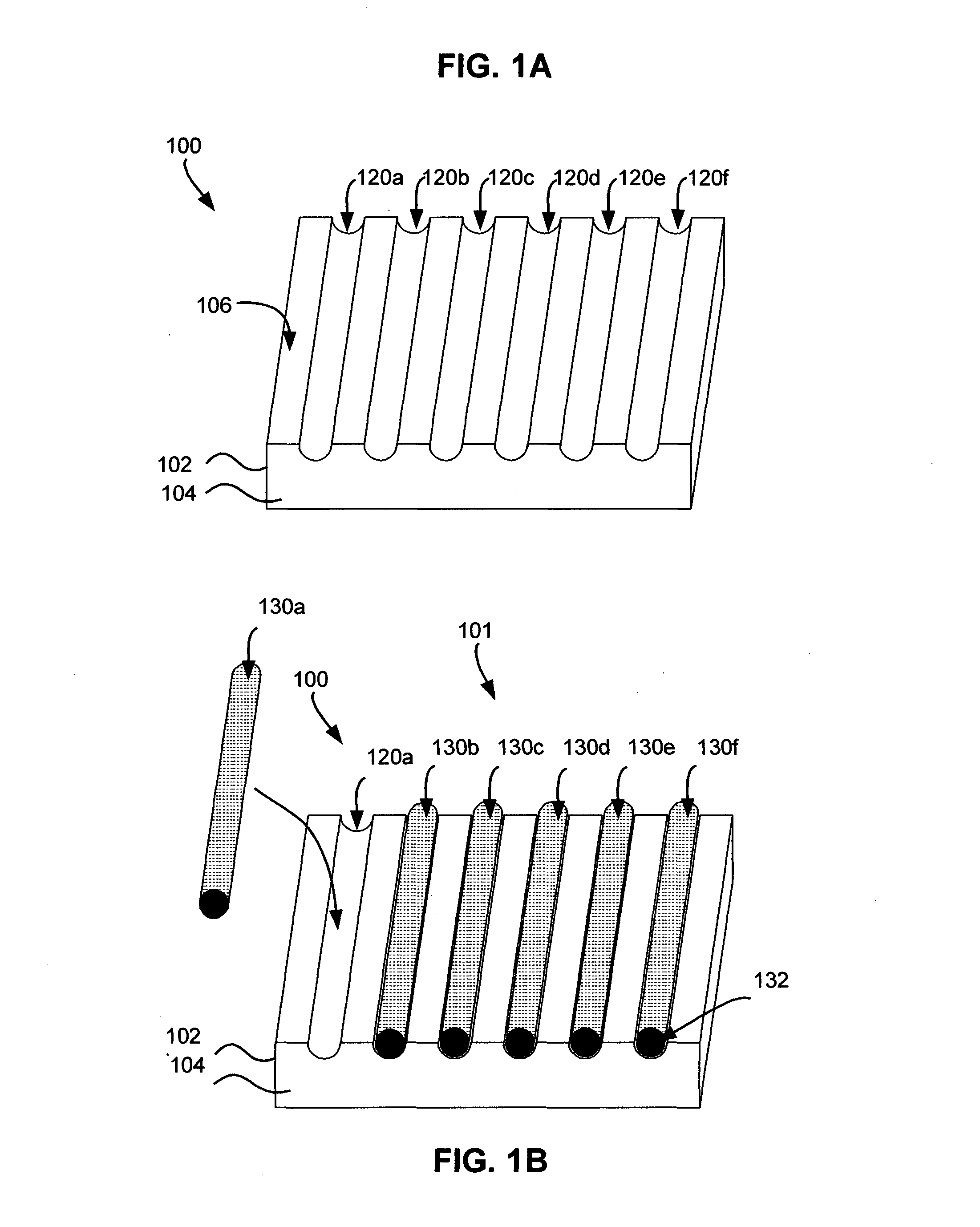
[0110]Method: A multiplex grooved matrix block 300d as disclosed herein was constructed from a protein gel (see FIG. 3D) and used for aligning the specimens by the urologist who collected the biopsies from 30 patients suspected of prostate cancer. Inclusion criteria for participants in the trial were based on clinical (positive DRE) or biochemical (PSA≧2.5 ng / ml) suspicion of prostate cancer. Up to 12 biopsy cores per patient (gauge 18) were collected with an ultrasound-guided biopsy gun in a single matrix and placed in a matrix 300d as shown in FIGS. 4A-4C. As shown in FIG. 4A, the biopsy core 400 was exposed at the tip of the needle 412 of the biopsy...


PUM


Abstract

A custom-made matrix suitable for receiving a tissue sample is described as well as the use thereof to obtain a multiplex histological preparation. The invention also relates to a multiplex biopsy array comprising tissue and / or cell samples arranged in a matrix material and to a method for the preparation of a multiplex biopsy array. Methods for preparing blocks of matrix material to be used in multiplex biopsy arrays are also described, as well as methods for loading biopsy samples in said blocks, and methods for treating and processing said blocks to form biopsy arrays. The biopsy arrays made using the block of matrix material can be used to prepare sections and slides for histological procedures, including quantitative analyses and parallel processing.

Description

CLAIM OF PRIORITY[0001]This application claims benefit or priority to U.S. provisional Patent Application Ser. No. 61 / 502,513 entitled “MATRIX AND METHOD FOR THE PREPARATION OF MULTIPLEX BIOPSY ARRAYS” filed Jun. 29, 2011 which is hereby incorporated by reference.CROSS-REFERENCES[0002]Not Applicable.FIELD OF THE INVENTION[0003]The invention relates to a custom-made matrix suitable for receiving a tissue sample and to the use thereof to obtain a multiplex histological preparation. The invention also relates to methods for preparing blocks of matrix material to be used in constructing multiplex biopsy arrays, methods for loading tissue samples in said blocks, and methods for treating and processing said blocks and arrays in preparation for analysis.BACKGROUND OF THE INVENTION[0004]The tissue sample may result from a needle biopsy (sometimes referred to as tru-cut biopsy), fine needle aspirate or an explant. A needle biopsy is a common medical test involving the removal with the aid of...

Claims


Application Information

Patent Timeline
IPC IPC(8): G01N33/543
CPCG01N33/5436G01N1/30G01N1/36G01N1/42A61K2300/00
Inventor MUSAT, SORIN
Owner LEAVITT MEDICAL
Learn More