
Apparatus for detecting tumor cells

a tumor cell and tumor cell technology, applied in the field of tumor cell detection apparatus, can solve the problems of large challenge of conventional tumor detection method, the failure of conventional cancer detection methods to identify about 40% of cancer patients who are in need of more or enhanced, and the failure of conventional cancer detection techniques such as x-ray imaging and nuclear magnetic resonance imaging to provide reliable information to the above critical applications, and achieve the effect of improving the signal-to-noise ratio

Pending Publication Date: 2014-03-13
ANPAC BIO MEDICAL SCI (LISHUI) CO LTD
View PDF0 Cites 8 Cited by
  • Summary
  • Abstract
  • Description
  • Claims
  • Application Information

AI Technical Summary

Benefits of technology

The apparatus enables early-stage cancer detection with high sensitivity and specificity, improving the accuracy of metastasis assessment and treatment efficacy, while reducing costs and equipment size.

Problems solved by technology

However, it is a huge challenge for conventional tumor detection methods to detect cancer earlier than symptomatically, or detect cancer at earlier stages of tumor metastasis.
For example, conventional methods fail to identify about 40% of cancer patients who are in need of more or enhanced therapies.
Conventional cancer detection techniques such as x-ray imaging and nuclear magnetic resonance (NMR) imaging fail to provide reliable information to the above critical applications.
Early detection and early systemic therapies will result in a declining death rate from cancer.
Metastasis, initiated by tumor cells transported through the circulation from the primary tumor to vital distant organs, is known to be the leading cause of cancer related deaths.
Present technique to separate and identify CTCs, on one hand, is quite labor intensive and expensive, and on the other hand lack accuracy and reliability.

Method used


Image

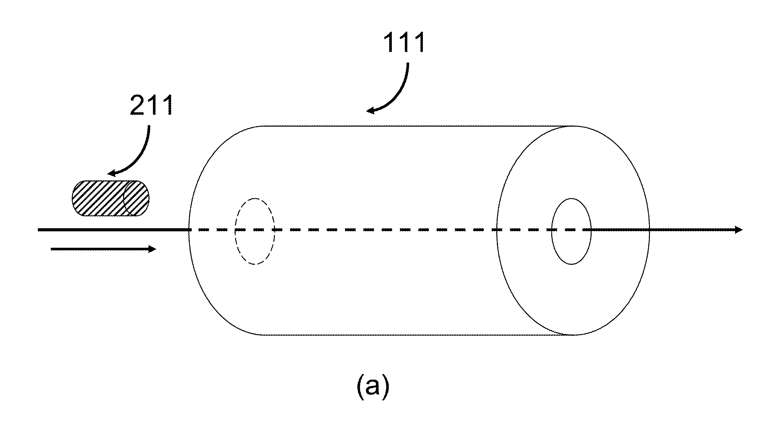
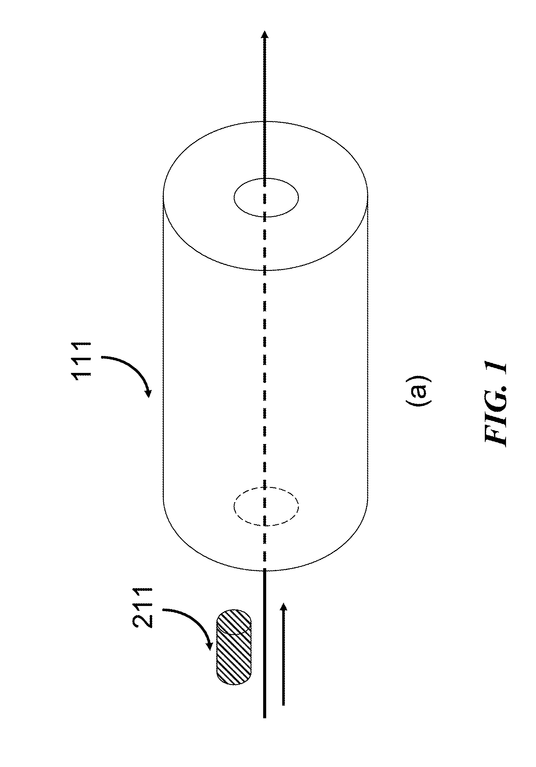
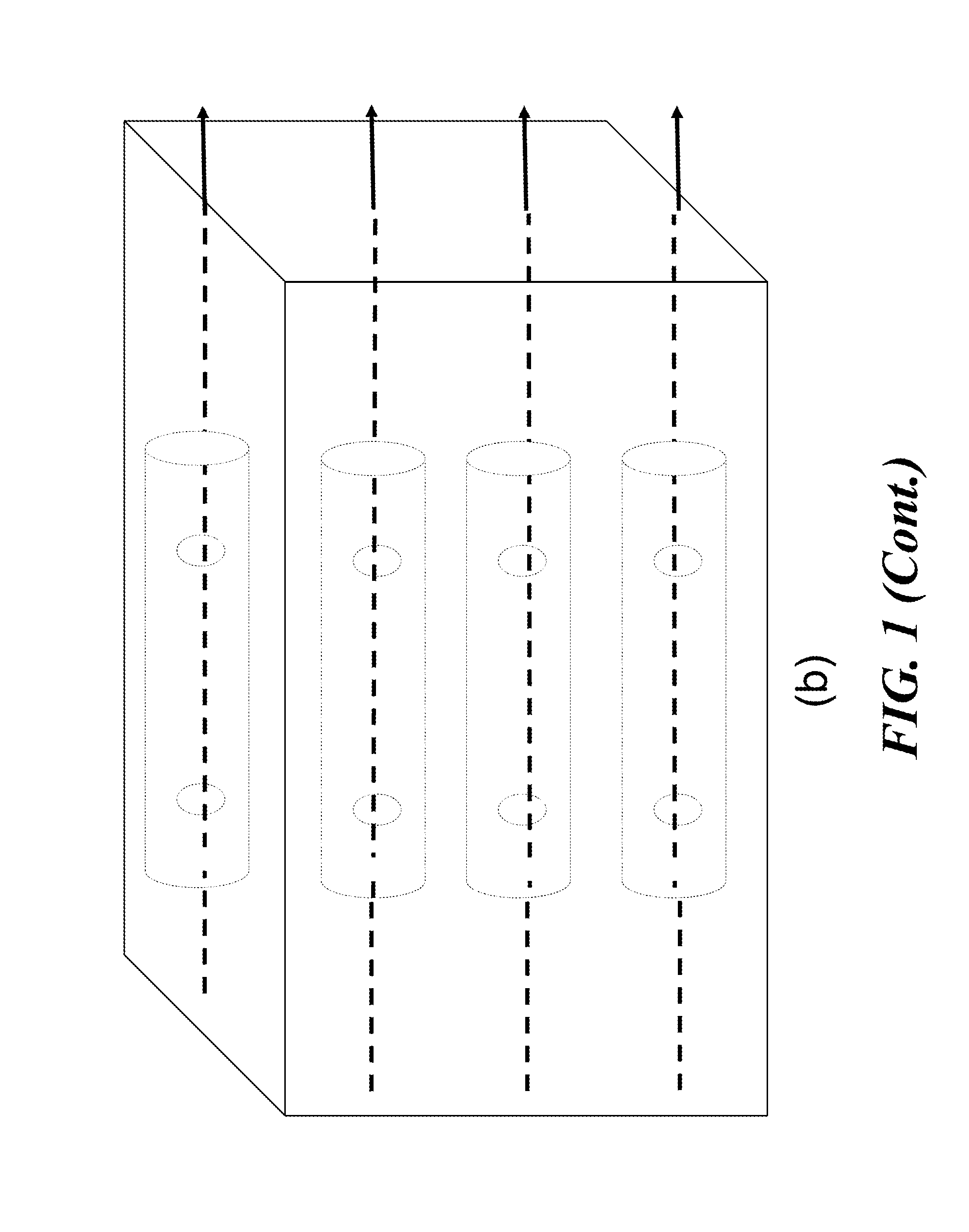
  • Apparatus for detecting tumor cells
  • Apparatus for detecting tumor cells
  • Apparatus for detecting tumor cells

Examples


Embodiment Construction

[0119]One aspect of the present invention relates to apparatus for detecting CTCs in a biological entity in vivo or in vitro (e.g., human being, an organ, a tissue, or cells in a culture). Each apparatus comprises a biological fluid delivering system, a pre-processing unit, a re-charging unit, a probing and detecting device, and a discharging unit. The apparatus is capable of measuring microscopic properties of a biological sample. By the constant pressure fluid delivery system, microscopic biological subjects can be delivered onto or into the pre-processing or diagnostic micro-device of the apparatus. Compared to traditional detection apparatus or technologies, the apparatus provided by this invention are advantageous in providing enhanced detection sensitivity, specificity, functionalities, and speed, with reduced costs and size. The apparatus can further include a biological interface, a probing controlling and data analysis circuitry, or a system reclaiming or treating medical w...


PUM


Abstract

Among others, the present invention provides apparatus for detecting circulating tumor cells, comprising a system delivery biological subject and a probing and detecting device, wherein the probing and detecting device includes a first micro-device and a first substrate supporting the first micro-device, the first micro-device contacts a biologic material to be detected and is capable of measuring at the microscopic level an electrical, magnetic, electromagnetic, thermal, optical, acoustical, biological, chemical, electro-mechanical, electro-chemical, electro-optical, electro-thermal, electro-chemical-mechanical, bio-chemical, bio-mechanical, bio-optical, bio-thermal, bio-physical, bio-electro-mechanical, bio-electro chemical, bio-electro-optical, bio-electro-thermal, bio-mechanical-optical, bio-mechanical thermal, bio-thermal-optical, bio-electro-chemical-optical, bio-electro-mechanical-optical, bio electro-thermal-optical, bio-electro-chemical-mechanical, physical or mechanical property, or a combination thereof, of the biologic subject.

Description

CROSS-REFERENCE TO RELATED APPLICATION[0001]This application claims priority to U.S. Application No. 61 / 482,900, filed on May 5, 2011, the contents of which are incorporated herein by reference in their entireties.BACKGROUND OF THE INVENTION[0002]A tumor is an abnormal growth of body tissue and can be cancerous (malignant) or noncancerous (benign). Tumors, particularly cancerous tumors, are a serious threat to human well-being and their detection in early stage is critical in order to obtain effective treatment or cure. However, it is a huge challenge for conventional tumor detection methods to detect cancer earlier than symptomatically, or detect cancer at earlier stages of tumor metastasis. For example, conventional methods fail to identify about 40% of cancer patients who are in need of more or enhanced therapies. It is also important to detect any early signs of spread in cancer following cancer treatments to assess effectiveness of the treatment, as well as if and what follow-u...

Claims


Application Information

Patent Timeline
Patent Type & Authority Applications(United States)
IPC IPC(8): A61B5/145A61B5/07
CPCA61B5/14546A61B5/14503A61B5/1451A61B5/14517A61B5/07B01L3/502707B01L3/502761G01N33/5091B01L2200/0668B01L2200/0663B01L2200/0647B01L2200/0652B01L2300/0645B01L2300/0636B01L2400/0487G01N15/1023A61B10/0041G01N33/487G01N33/54366G01N15/01G01N2015/1024C12Q1/68G01N1/34B01L2300/0887A61B5/14528A61B2562/028A61B2562/12B01L2300/0681B01L2300/163G01N1/4077G01N15/1484G01N33/574G01N2001/4088G01N2015/1006G01N2015/1486B01L3/502715B01L3/50273B01L2300/0627B01L2300/0858B01L2300/123
Inventor YU, CHRIS C.DU, XUEDONGYU, HE
Owner ANPAC BIO MEDICAL SCI (LISHUI) CO LTD
Learn More