Dna-origami-based standard

a technology of dna-origami and standard, applied in the field of dna-origami-based standard, can solve the problems of limited quantity analysis using such microscopy techniques, limited availability of standardized samples, and large size dimensions, and can only be combined with great difficulty with the requirements on the molecular scal

Inactive Publication Date: 2014-02-27
TECH UNIV BRAUNSCHWEIG
View PDF1 Cites 11 Cited by
  • Summary
  • Abstract
  • Description
  • Claims
  • Application Information

AI Technical Summary

Problems solved by technology

However, quantitative analysis using such microscopy techniques is limited in that there are only a few methods which allow calibration of these measuring devices, such as microscopes.
Particularly the provision of standardized samples is limited, especially in submicrometer ranges right up to the ranges of super-resolution imaging and FRET.
Top-down lithographic approaches can attain the required size dimensions, but can be combined only with great difficulty with the requirements on the molecular scale.
In addition, such approaches are usually not biocompatible or optically compatible and influence, in particular, also the properties of the labeling molecules, such as the fluorescent dyes used in the area of fluorescence microscopy.
Chemical and macromolecular approaches, as used in bottom-up approaches, can form regular structures in the required size, but there is then a problem in the structural and stoichiometric determination in the ranges relevant to microscopy, since individual nano-objects such as fluorescent dyes cannot be placed at the relevant intervals.
The quenching effect does not allow quantitative analysis.
However, the disadvantage here is that the excitation light intensity actually arriving in the sample is not measured.
In some cases, such a measurement is not even possible owing to specific peculiarities of the method, for example in the case of TIRF excitation.
Moreover, the light-sensitive detector measures the integral intensity, but the intensity of the excitation light is subject to great heterogeneity, which is not taken into account.
However, a disadvantage thereof is that the beads usually do not contain a defined number of dyes.
Moreover, the dye molecules are present in the beads in an unordered manner, and so interactions between the individual dye molecules occur.
The sensitivity of a microscope cannot be precisely determined owing to the relatively large signal heterogeneity.
It is also no longer possible to calibrate the sensitivity to the number of detectable dyes, since not all dyes are equally bright.
Similarly, it is not possible to exactly deduce an excitation output prevailing at a site.
However, the barcode probes described therein are not useful for calibration of measuring derives, like microscope.
In particular, the barcode probes do not allow any calibration for quantitative analysis.

Method used


Image

  • Dna-origami-based standard
  • Dna-origami-based standard
  • Dna-origami-based standard

Examples


example 1

Brightness Standards Based on DNA Origami

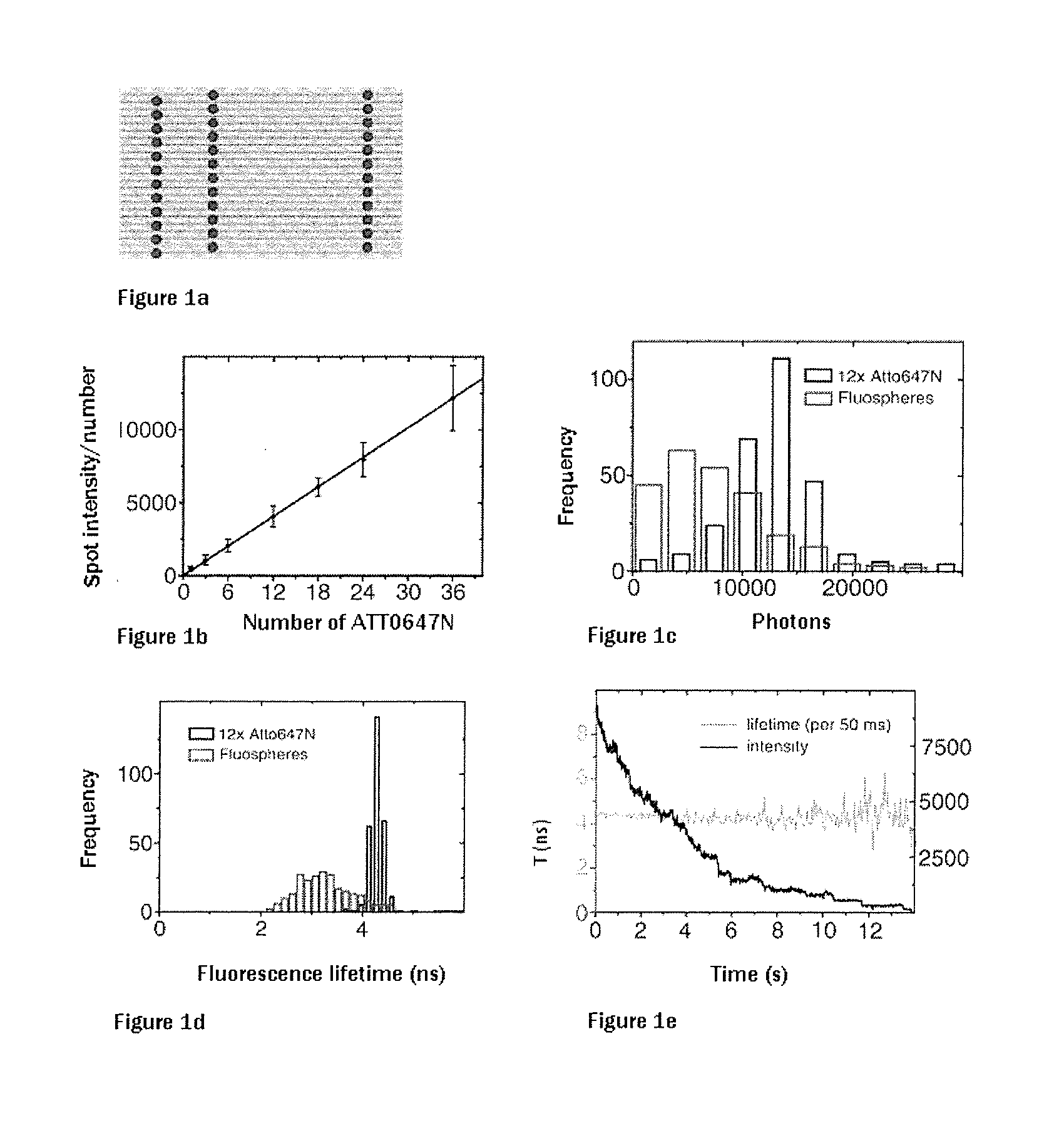
[0058]The ATTO647N-labeled short DNA segments were used in the self-assembly of the DNA origami. FIG. 1a shows a corresponding diagram of a rectangular DNA origami having 36 fluorophore positions. FIG. 1b shows the analysis of the spatially integrated photon number based on the number of labeling molecules. The linear direct dependence of the number of photons as a measure of the brightness of the number of incorporated fluorophores can be clearly seen. To this end, DNA origami having 12, 24 and 36 ATTO647N molecules were used. It is clear that there is no discernible self-quenching which leads to a reduction in the photons per spot. In contrast, experiments with commercially available beads in which the fluorophores are randomly distributed show that self-quenching occurs (FIG. 1c). Furthermore, the lifetime of the fluorescence in the case of the DNA origami sample is very homogeneous in contrast to the commercially used beads (FIGS. 1d and ...

example 2

Standards for STED Microscopy

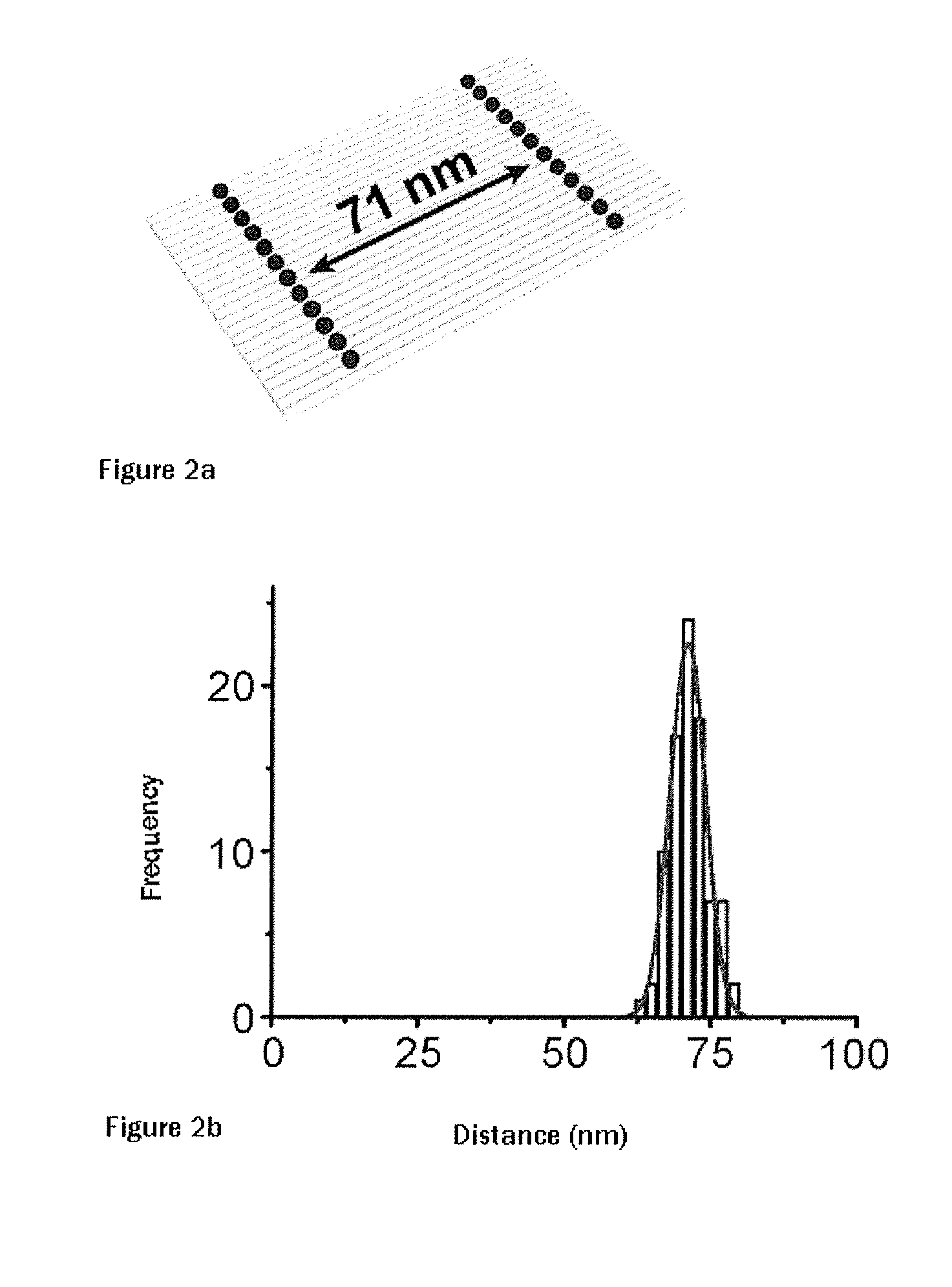
[0060]STED (stimulated emission depletion) was the first super-resolution microscope technology which breached the diffraction limit. DNA origami rulers were prepared here for both pulsed and continuous STED. To this end, corresponding rectangular origami were prepared with a distance of 71 nanometers between the two lines composed of, in each case, 12 ATTO647N molecules (see FIG. 2a). Said DNA origami were immobilized on polylysine-coated cover slips and covered with a polymer layer. Using STED technology, it was possible to resolve the interval between the two lines composed of, in each case, 12 molecules, and it was possible by means of STED microscopy to determine the distance between the two lines to 71±3 nm, as shown in FIG. 2b. Using STED with pulsed excitation, it was also possible to resolve lines at an interval of 44 nanometers. Similar results could be achieved with Alexa 488 fluorophores (data not shown).

example 3

Standards for Ultra-High Resolution Imaging

[0061]The resolution of super-resolution microscopy below the diffraction limit is normally limited by (i) photobleaching, (ii) the measured photon numbers in an “on state” and the on / off cycle or simply because of the stability of the structure. Here, rectangles having two ATTO647N molecules at intervals of 6, 12 and 18 nm were designed in DNA origami (see FIG. 3a). Said DNA origami were immobilized with 5 biotin-labeled strands. To avoid limitation by the number of photons, the fluorescence of the dyes was captured until photobleaching. Subsequently, the positions of the individual dyes were determined by subtracting the point spread function of the longer-lived dye from the point spread function before the first photobleaching step. The individual molecules were localized in reverse order of the photobleaching and the intensity distribution of the second molecule was subtracted from the first part of the transition. By way of example, it...


PUM

PropertyMeasurementUnit
distanceaaaaaaaaaa
pHaaaaaaaaaa
wavelengthaaaaaaaaaa

Abstract

Arrays which utilize labeling molecules for calibrating a measuring device, such as microscopes, have a first structure based on a DNA origami as a calibration sample, wherein the DNA origami is formed into a predetermined structure by short DNA segments. The DNA origami is optionally present in an arranged manner on a support, wherein a number of short DNA segments which form the predetermined structure include a labeling molecule. Optionally, the array can have at least a second structure based on a DNA origami, different from the first structure, as a calibration sample. The array allows quantification of the labeling molecules on the basis of the number of photons per unit time.

Description

[0001]The present invention is directed to standards suitable for calibrating measuring devices, more particularly microscopes. More precisely, the present invention relates to arrays for calibrating a measuring device using labeling molecules, wherein said array has a first structure based on a DNA origami as a calibration sample and wherein the DNA origami is formed into a predetermined structure by means of short DNA segments and is optionally present in an arranged manner on a support, wherein a predetermined number of the short DNA segments which can form the predetermined structure of the DNA origami has a predetermined number of a labeling molecule. Optionally, the array can have at least a second structure based on a DNA origami, different from the first structure, as a calibration sample. The array for measuring-device calibration is particularly suited for quantifying measurement signals, more particularly it allows quantification of the labeling molecules on the basis of ...

Claims


Application Information

Patent Timeline
Patent Type & Authority Applications(United States)
IPC IPC(8): G01N21/17
CPCG01N21/17G01N21/6458
Inventor TINNEFELD, PHILIPSCHMIED, JUERGENHOLZMEISTER, PHIL
Owner TECH UNIV BRAUNSCHWEIG
Why Eureka
Try Eureka