High-Speed Tumor Segmentation System

a tumor and high-speed technology, applied in the field of tumor segmentation in medical imaging, can solve the problems of reducing the accuracy of dose placement, affecting the accuracy of tumor dose placement, and time-consuming process of tumor volume segmentation, and achieve the effect of fast automatic segmentation

Active Publication Date: 2013-10-10
WISCONSIN ALUMNI RES FOUND
View PDF0 Cites 7 Cited by
  • Summary
  • Abstract
  • Description
  • Claims
  • Application Information

AI Technical Summary

Benefits of technology

[0009]The present invention provides an extremely fast automatic segmentation technique for use with radiation therapy or other applications, and providing real-time medical imaging. The segmentation technique works by dividing the data into multiple regions based on data value similarity. Selected regions are then identified as tumors using an area filtering exploiting rough a priori knowledge about the tumor size optionally augmented by tumor location. Rapid morphological operators are used to generate the multiple regions at speeds allowing real-time tumor motion and deformation correction.
[0011]It is thus a feature of at least one embodiment of the invention to provide a segmentation approach that permits real-time correction of the radiation beam during a treatment session. Segregating data according to data values may be performed in a one-pass rapid calculation making the tumor segmentation a simple selection among these regions.
[0021]It is thus a feature of at least one embodiment of the invention to focus the segmentation process on information about the tumor to provide greatest accuracy in the tumor segmentation.
[0023]It is thus a feature of at least one embodiment of the invention to provide isolated regions using extremely fast morphological processing.
[0025]It is thus a feature of at least one embodiment of the invention to provide a robust and rapid method for refining the identified regions.
[0029]It is thus a feature of at least one embodiment of the invention to provide a flexible output format for different radiation beam correction systems.

Problems solved by technology

The process of segmenting tumor volume can be time-consuming, requiring a healthcare professional to draw boundary lines on multiple image “slices” that together describe a volume of tissue to be treated.
During the radiation treatment, ability to precisely control the dose to the tumor is significantly limited by uncertainties in the position and size of the tumor.
“Fractionation”, which breaks the radiation therapy into multiple sessions on different days, each delivering a fraction of the total desired dose, may be used to increase the susceptibility of the tumor cells and allow healthy cells to repair between fractions, but can reduce the accuracy of dose placement because of tumor position changes between fractionation sessions.
Further, the tumor size may change over time (regression) as a result of the treatment itself.
Even during a particular treatment session, uncertainty in the tumor position can result from periodic organ movements such as those of the heart and lungs or aperiodic motion such as bladder filling.
The process of segmenting the tumor in each of these images is extremely time-consuming and prone to error.
Manual and semiautomatic segmentation is much too slow to exploit this ability.
Unfortunately, automatic segmentation techniques such as thresholding, region growing, clustering, and neural networks, have failed to achieve sufficiently accurate or rapid segmentation for real-time tumor segmentation.

Method used


Image

  • High-Speed Tumor Segmentation System
  • High-Speed Tumor Segmentation System
  • High-Speed Tumor Segmentation System

Examples


Embodiment Construction

Hardware

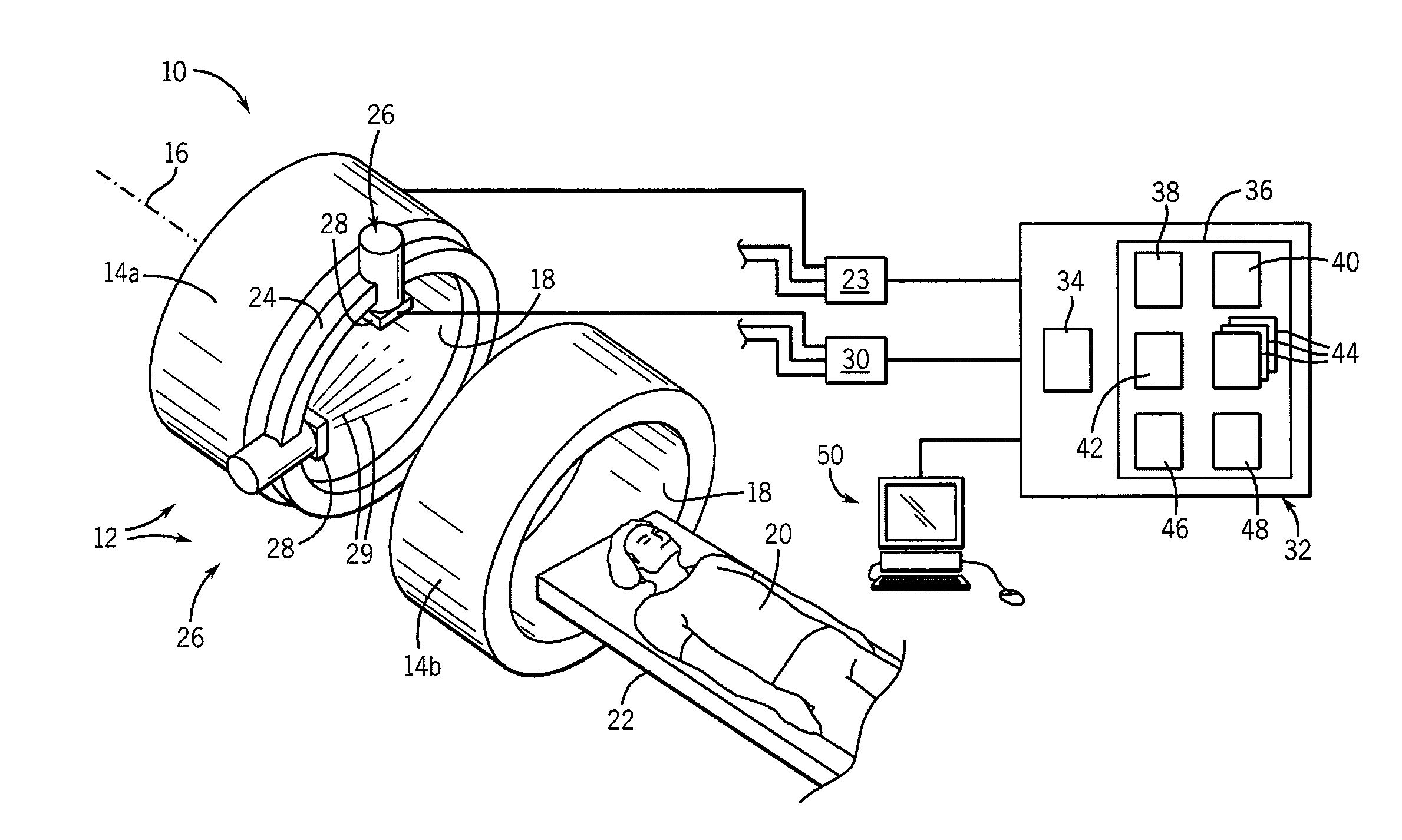
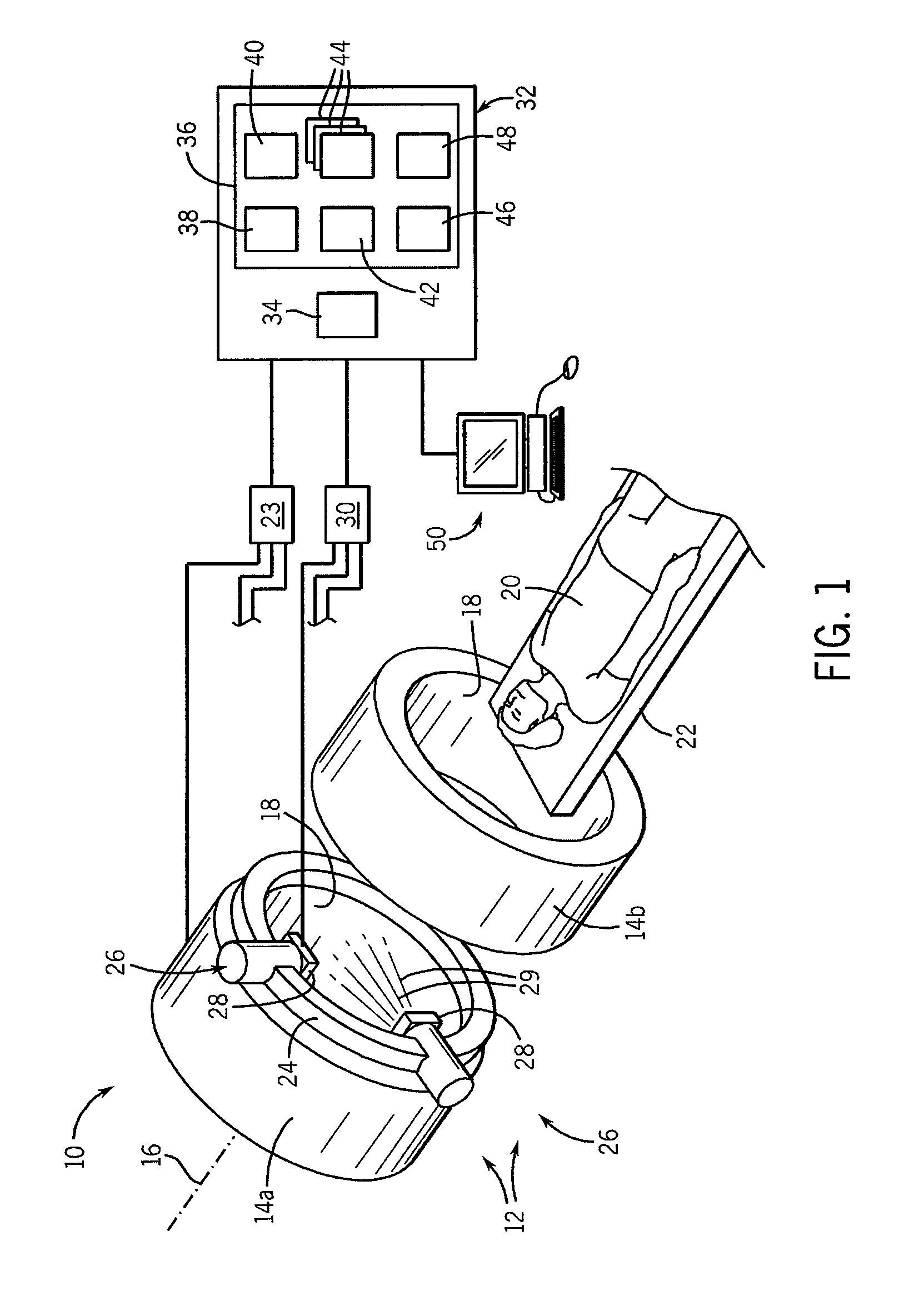
[0036]Referring now to FIG. 1, a combination imaging radiotherapy machine 10 may provide, for example, an MRI magnet assembly 12 having two toroidal magnet elements 14a and 14b spaced along an axis 16 and having aligned bores 18 sized to admit a patient 20 supported on a patient table 22. The magnet elements 14a and 14b may hold a superconducting BO electromagnet and one or more gradient and RF coils as is understood in the art of magnetic resonance imaging. The gradient and RF coils may communicate with an MRI subsystem 23 providing power amplifiers and the like as is also understood in the art.

[0037]A radiation therapy gantry 24 may be positioned between the magnet elements 14a and 14b, and may hold one or more radiation delivery systems 26 positioned to rotate about the axis 16 and direct radiation beams 29 toward the patient 20 when the patient 20 is positioned within the MRI magnet assembly 12. Each radiation delivery system 26 may include an electronically controllable...


PUM


Abstract

A system for automatic segmentation of tumor tissue, useful for motion correction during radiotherapy using real-time imaging, identifies multiple regions based on the values of data and then identifies the tumors within the regions based on a priori knowledge about tumor size and / or location. The regions may be refined with robust and fast morphological operations, providing segmentation at speeds commensurate with the motion to be corrected.

Description

BACKGROUND OF THE INVENTION[0001]The present invention relates to tumor segmentation in medical imaging with applications in radiation therapy, surgery and chemotherapy, and, in particular, to a method and apparatus for real-time tracking of tumor location for improved radiation therapy.[0002]External beam radiation therapy treats tumor tissues by directing a beam of high-energy radiation into the patient and through the tumor, exploiting increased sensitivity of rapidly dividing tumor cells to radiation damage. Radiation, and radiotherapy, as used herein, will generally be understood to include high-energy electromagnetic radiation, as well as particles such as protons.[0003]The success of external beam radiation therapy is strongly linked to the delivered dose, the latter which is constrained by the need to protect tissue and organs near the tumor. Damage to healthy tissue can be reduced by directing the radiation beam along a number of different axes that all intersect at the tum...

Claims


Application Information

Patent Timeline
Patent Type & Authority Applications(United States)
IPC IPC(8): A61N5/00
CPCA61N5/1049A61N5/1067G06F19/321A61N2005/1061A61N2005/1055G16H30/20G16H30/40
Inventor CHEBROLU, VENKATA VEERENDRANADHPALIWAL, BHUDATT R.
Owner WISCONSIN ALUMNI RES FOUND
Why Eureka
Try Eureka