
Probe for diagnosis and treatment of muscle contraction dysfunction

a muscle contraction and muscle technology, applied in the field of probes, can solve the problems of insufficient design of adhesive surface electrodes that are commonly used for emg recordings of other skeletal muscles, ineffective adhesion in moist mucus membrane environment, and only useful surface electrodes

Inactive Publication Date: 2013-06-13
QUEENS UNIV OF KINGSTON
View PDF10 Cites 29 Cited by
  • Summary
  • Abstract
  • Description
  • Claims
  • Application Information

AI Technical Summary

Benefits of technology

The invention is a probe that has several advantages over existing probes. It is less likely to pick up unwanted signals and is more comfortable to use. It also doesn't change the way muscles behave or move during tasks that increase pressure.

Problems solved by technology

However, surface electrodes are generally only useful for recording the activity from muscles close to the skin's surface.
Despite the possible importance of the superficial PFMs in women with PPM dysfunction, commercially available intravaginal probes do not record activity from these muscles.
As a result, design of adhesive surface electrodes that are commonly used for EMG recordings of other skeletal muscles is not appropriate.
For example, for EMG of an arm muscle, an adhesive electrode can adhere to the skin of the arm, but such adhesion is ineffective in a moist mucus membrane environment.
Drawbacks with currently available technology include that most probes use a monopolar electrode configuration with either large circumferential electrodes encircling the probe or one electrode on each side of the probe.
Currently available probes have electrode configurations that do not allow for different pelvic floor muscles on each side of vaginal canal (levator hiatus) to be investigated or presented separately to the patient for assessment or biofeedback training.
The probes, being rather large, can also be uncomfortable, especially if they are used to record activity when the user changes positions or performs a functional activity (Brown, C., 2007, Reliability of Electromyography Detection Systems for the Pelvic Floor Muscles, retrieved from http: / / hdl.handle.net / 1974 / 948).
In addition, there is little to no control over where the electrodes sit with respect to the location of the pelvic floor muscles in a given user when the probe is inserted into the vagina.
Electromyographic (EMG) signals can also be contaminated by motion artifact, affecting the signal's validity.
The moist environment of the vagina does not allow for electrodes to be adhered to the vaginal wall, thus most intravaginal electrodes are prone to motion artifact.
Given the rigid nature of a vaginal probe, it is suspect to much movement if users perform functional activities or tasks.
In particular, electrodes mounted on intravaginal probes are subject to motion artifact during actions such as coughing, laughing or sneezing, in which an abrupt and strong increase in intra-abdominal pressure generates a caudal force at the level of the probe.
In sum, commercially available intravaginal probes possess deficiencies in their design such as problems with probe geometry, electrode size, location, and / or configuration.

Method used


Image

  • Probe for diagnosis and treatment of muscle contraction dysfunction
  • Probe for diagnosis and treatment of muscle contraction dysfunction
  • Probe for diagnosis and treatment of muscle contraction dysfunction

Examples


example 1

[0092]A study was performed to determine the reliability and validity of Probe 1 of the invention when recording surface EMG from the PFMs in healthy women. Probe 1 was also compared to a commonly used electrode (Femiscan™; surface area 1.75 cm2 each). The Femiscan™ device was re-wired to record differential configurations from the right and left PFMs separately since this is a more appropriate way to record such muscle activation.

[0093]Reliability refers to between-trial reliability for the probe. Between-day reliability is not expected to be high for any EMG data since there is, among other factors, inherent variation in electrode position relative to active muscle fibers. Validity refers to the effect of the hip adductor (Add) and external rotator (ER) contractions on the signal recorded at the PFMs. In this case we were particularly interested in determining whether the recorded EMG signals come from the PFMs or represent crosstalk from nearby muscles.

[0094]Twenty healthy nullip...

example 2

Determination of Motion Artifact

[0107]A study was performed to determine whether EMG recordings made using the novel probe described herein have less motion artifact contamination than the Femiscan™ electrode, a commercially available electrode reconfigured to incorporate two differential EMG channels (one on each side of the vaginal wall) using stainless steel bars mounted on a cylindrical probe.

Methods:

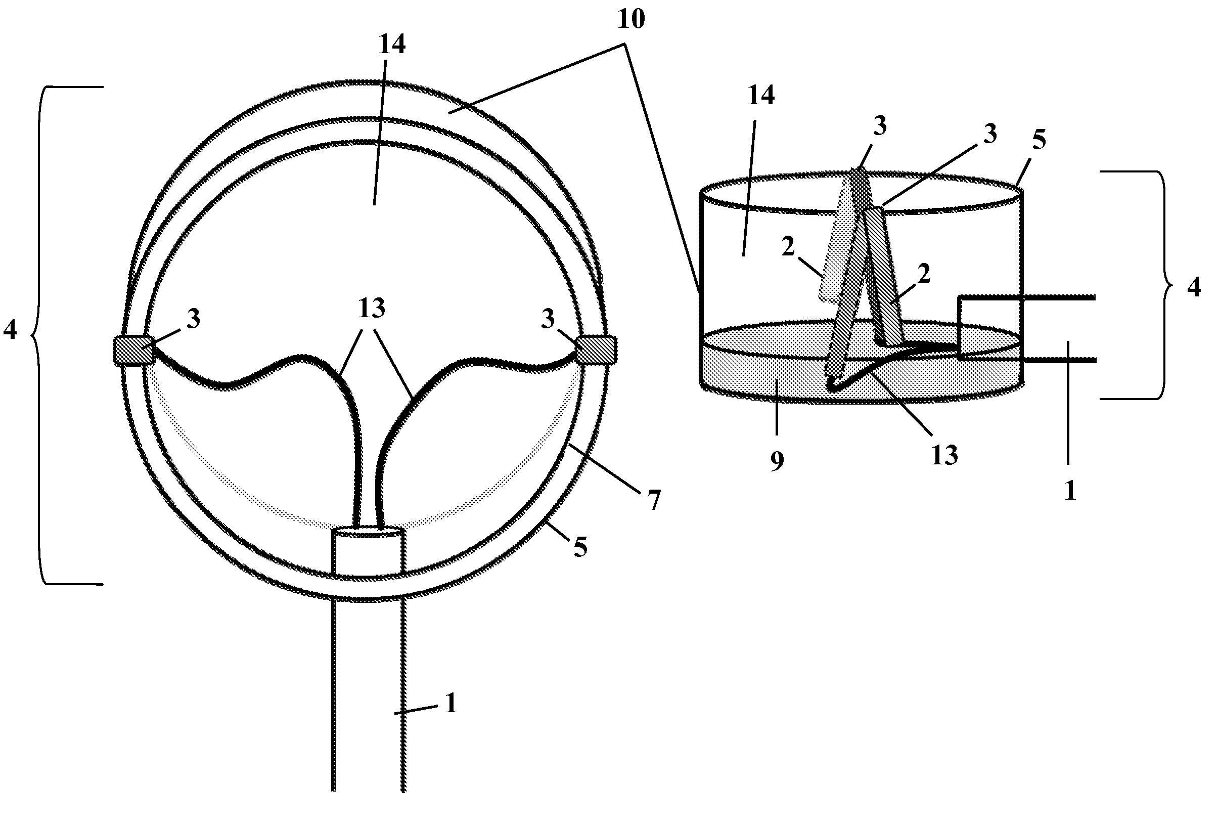
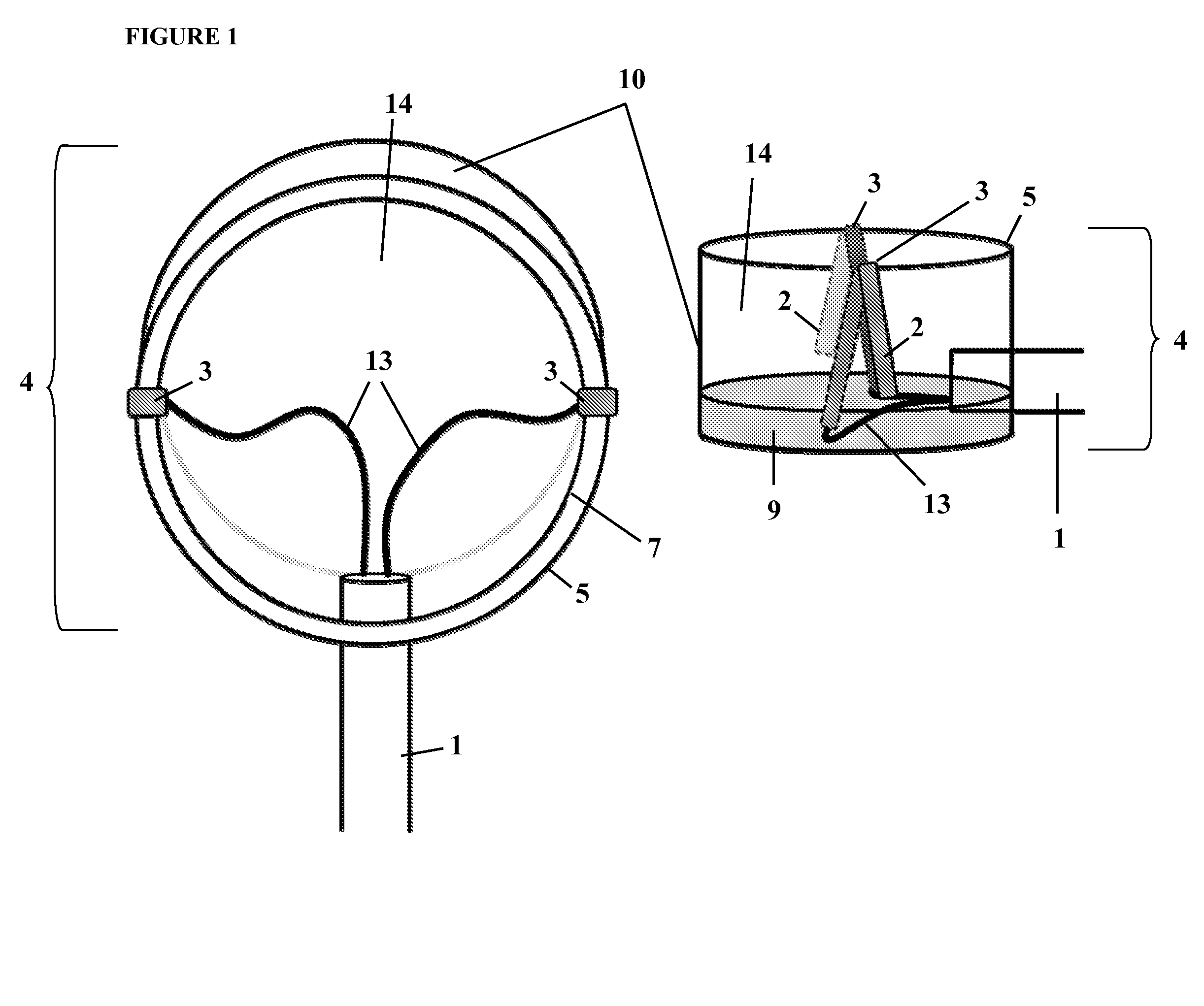
[0108]Eighteen healthy continent women with no signs of pelvic floor muscle dysfunction (such as urinary or fecal incontinence, pelvic pain disorders, or low back pain) were recruited from the Kingston (Ontario, Canada) community. Each participant performed ten repetitions of a maximal effort coughing task in the standing position with both the Femiscan™ probe and Probe 1 of the invention (see FIG. 1) in situ, with the probes tested in random order. EMU data were recorded from both sides of the vaginal wall using Delsys™ AMT-8 pre-amplifiers (bandwidth 20-450 Hz, input impedance >10...

example 3

Determination of Crosstalk

[0116]A study was performed on three volunteers (healthy, nulliparous women) to determine whether EMG recordings made using Probe 4 have crosstalk contamination from the obturator internus muscle (Exemplar data are presented in FIG. 1). EMG data were recorded from PFMs during a contraction of the hip external rotators, which should elicit obturator internus activity but not pelvic floor muscle activity. The following data were recorded: pelvic floor muscle EMG data were recorded using fine wire electrodes located in the right pelvic floor muscle (gold standard) (top panel of FIG. 11); obturator internus EMG data were recorded using fine wire electrodes placed in the right obturator internus muscle (second panel from top in FIG. 11); pelvic floor muscle EMG data were recorded simultaneously using Probe 4 (bottom two panels in FIG. 11; third panel from top shows data recorded with the probe located on the left side of the vagina, and the bottom panel shows da...


PUM


Abstract

A novel probe for recording EMG signals from muscles, in particular intravaginal signals from the pelvic floor muscles (PFM), is provided herein. The probe includes an insertion end having a suction head forming a vessel open at the top with attached electrodes and a distal end for attachment to a means of providing suction and an amplifier.

Description

CROSS-REFERENCE TO RELATED APPLICATIONS[0001]This application claims priority to U.S. provisional application No. 61 / 375,613 filed Aug. 20, 2010, the entire contents of which are hereby incorporated by reference.FIELD OF THE INVENTION[0002]The present invention relates to a novel probe for recording EMG signals from muscles, in particular intravaginal signals from the pelvic floor muscles (PFMs).BACKGROUND[0003]Electromyography (EMG) is a tool used to record electrical voltages induced through ion shifts that occur when a muscle contracts. The arrival of an action potential at the neuromuscular junction triggers changes in muscle cell membrane permeability, eventually leading to the formation of muscle fiber action potentials. An EMG signal is a recording of all muscle fiber action potentials located within the vicinity of the detection surfaces of the particular electrodes used and is a convenient way to determine the timing and extent of neuromuscular activation. Surface EMG is th...

Claims


Application Information

Patent Timeline
IPC IPC(8): A61B5/0488A61B5/296
CPCA61B5/04882A61B5/0492A61B5/202A61B5/6879A61B5/0488A61B5/6852A61B5/4337A61B5/391A61B5/296A61B5/389
Inventor MCLEAN, LINDA B.YOUNG, ROY A.
Owner QUEENS UNIV OF KINGSTON
Learn More