Three-dimensional scaffolds, methods for fabricating the same, and methods of treating a peripheral nerve or spinal cord injury

a three-dimensional scaffold and scaffolding technology, applied in the field of three-dimensional scaffolds, can solve the problems of limited treatment of spinal cord injury, regeneration failure, and failure of axons to regenerate spontaneously, and achieve the effects of long distance regeneration, robustness, and extensive axonal regeneration

Active Publication Date: 2013-05-02
RENESSELAER POLYTECHNIC INST +1
View PDF5 Cites 34 Cited by
  • Summary
  • Abstract
  • Description
  • Claims
  • Application Information

AI Technical Summary

Benefits of technology

This patent describes a method for making a three-dimensional scaffold that can be used to treat various medical conditions. The scaffold is made up of highly-aligned fibers that are electrospun into a layer and then shaped into a scaffold without disturbing the alignment of the fibers. The scaffold has a base layer that is curved along one or more axes and is made of a polymer film that is porous. The scaffold is designed to provide a surface for cells to adhere and proliferate, while also providing mechanical stability and support. The scaffold has a unique shape or form, which can influence or delimit a three-dimensional shape or form assumed by a population of proliferating cells. The scaffold can also contain a therapeutic agent that can be effective in treating or preventing medical conditions. The amount of therapeutic agent needed for treatment can be determined based on the individual's condition and prescribed by a physician or veterinarian.

Problems solved by technology

One major factor that limits treatment of spinal cord injury is the failure of axons to regenerate spontaneously after central nervous system (CNS) injury.
Regeneration failure can be partially attributed to a limited intrinsic axon growth capacity of adult neurons.
In addition to a limited growth capacity, an inhospitable extracellular environment hinders regeneration.
However, harvesting autografts is limited by tissue availability and donor site morbidity, and the alternative acellularized allografts fail to support axonal regeneration.
Nevertheless, poor survival limits the potential of cellular therapies, and neurotrophin administration requires precise timing, dose, and spatial distribution.

Method used


Image

  • Three-dimensional scaffolds, methods for fabricating the same, and methods of treating a peripheral nerve or spinal cord injury
  • Three-dimensional scaffolds, methods for fabricating the same, and methods of treating a peripheral nerve or spinal cord injury
  • Three-dimensional scaffolds, methods for fabricating the same, and methods of treating a peripheral nerve or spinal cord injury

Examples


example 1

Aligned Microfibers Foster Efficient Dorsal Root Ganglia Neurite Growth

[0114]Polymer microfiber substrates were fabricated by electrospinning an 8% PLA solution onto 20 μm thin PLA films (FIG. 1) (Wang et al., 2009). Randomly oriented fibers were deposited on films by electrospinning PLA onto a stationary target; a rotating target was used to make aligned fiber substrates as described in our previous work (Wang et al., 2006). Fiber diameter and alignment were analyzed from scanning electron micrographs (FIGS. 2D and 2E and FIGS. 3A and 3C). Fiber diameter ranged between 1.2 and 1.6 μm. Only 4% of random fibers were found to fall within ±4° C. of the median fiber orientation (FIG. 3B), whereas 97.3% of aligned fibers fell within ±4° of the median orientation (FIG. 3D).

[0115]Dorsal root ganglia (DRG) isolated from P4 rat pups were cultured on film, random, and aligned fiber substrates for 5 days in serum free media (n=6). DRG cultured on film and random fiber substrates projected neur...

example 2

Microtopography Supports Host Tissue Integration and Gap Closure

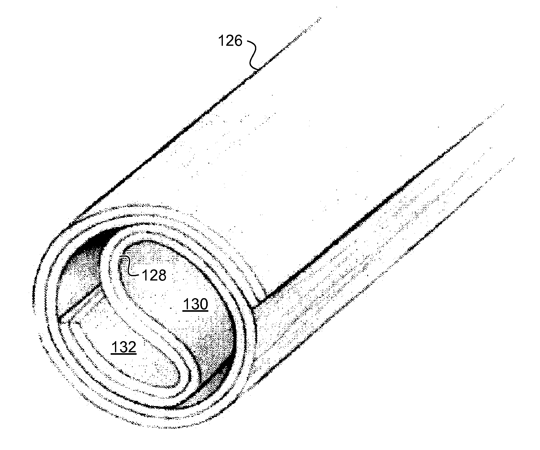
[0116]Guidance conduits were fabricated from film, random, and aligned fiber 2D substrates rolled into conduits (FIGS. 1C-1F and FIG. 3H). Importantly, conduit fabrication did not perturb fiber alignment. 93.3% of fibers fell within ±4° C. of the median orientation post-fabrication (FIG. 3F). An unambiguous, complete transection model of rat spinal cord injury was used to test the hypothesis that aligned microfibers promote axonal regeneration after CNS injury (FIG. 13). A 3 mm gap was created in the spinal cord (thoracic 9) of adult rats, and immediately after, film, random, or aligned fiber conduits filled with a fibrin gel were grafted into the gap (FIG. 4). Animals from each group were perfused 1 (n=3), 2 (n=3), and 4 weeks (n=7) following injury / implantation. The results from the 4 week time point were gathered in two independent experiments of n=3 and n=4 for each group.

[0117]Cresyl violet and DAPI staining were u...

example 3

Aligned Fibers Promote Robust, Long Distance Axonal Regeneration

[0119]Horizontal sections were immunolabeled for neurofilament (RT97) and serotonin (5HT) to examine axonal regeneration after complete spinal cord transection. Neurofilament section overviews demonstrated robust rostrocaudal axonal regeneration into aligned fiber conduits. One week after implantation, all groups displayed a similar pattern of RT97 staining (FIGS. 8A-8C). However, by 2 weeks neurofilament staining demonstrated robust rostrocaudal growth into aligned fiber conduits (FIG. 8F) that further elongated by 4 weeks (FIG. 8I). This response was observed exclusively from the rostral spinal cord. In random fiber conduits, individual RT97+ axons entered the grafts at 2 weeks (FIG. 8E), and a modest rostrocaudal response was observed by 4 weeks (FIG. 8H). In film conduits, very few axons were present at the interface at 2 weeks (FIG. 8D), and at 4 weeks sparse RT97+ axons were seen inside thin tissue strands (FIG. 8...


PUM

PropertyMeasurementUnit
Lengthaaaaaaaaaa
Lengthaaaaaaaaaa
Fractionaaaaaaaaaa

Abstract

One aspect of the invention provides a three-dimensional scaffold including at least one layer of highly-aligned fibers. The at least one layer of highly-aligned fibers is curved in a direction substantially perpendicular to a general direction of the fibers. Another aspect of the invention provides a method for fabricating a three-dimensional scaffold. The method includes: electro spinning a plurality of fibers to produce at least one layer of highly-aligned fibers and forming the at least one layer of highly-aligned fibers into a three-dimensional scaffold without disturbing the alignment of the highly-aligned polymer fibers. A further aspect of the invention provides methods for using a three-dimensional scaffold to treat nerve or spinal cord injury.

Description

CROSS-REFERENCE TO RELATED APPLICATION[0001]This application claims the benefit of U.S. Provisional Patent Application Ser. No. 61 / 320,088, filed Apr. 1, 2010, the contents of which is incorporated herein by reference in its entirety.STATEMENT OF RIGHTS TO INVENTIONS MADE UNDER FEDERALLY SPONSORED RESEARCH[0002]This work was supported by grant no. NINDS R21NS62392 and NICHD R15HD61096 from the National Institutes of Health. The U.S. Government has certain rights in this invention.BACKGROUND OF THE INVENTION[0003]In the United States, it is estimated that 11,000 individuals sustain spinal cord injuries each year. One major factor that limits treatment of spinal cord injury is the failure of axons to regenerate spontaneously after central nervous system (CNS) injury. Regeneration failure can be partially attributed to a limited intrinsic axon growth capacity of adult neurons. Indeed, augmenting the growth potential of centrally projecting axons via peripheral conditioning or other cAM...

Claims


Application Information

Patent Timeline
IPC IPC(8): A61F2/00
CPCA61F2/04A61F2/0063A61L27/383A61L27/3834A61L27/54A61L2300/254A61L2300/414A61L2300/416A61L2430/32A61L2430/38D01D5/0076D06M16/003A61L27/18C08L67/04A61F2/00
Inventor HURTADO, ANDRESGILBERT, RYAN J.WANG, HAN B.CREGG, JARED M.MULLINS, MICHAEL E.OUDEGA, MARTIN
Owner RENESSELAER POLYTECHNIC INST
Why Eureka
Try Eureka