
Simulated, representative high-fidelity organosilicate tissue models

a tissue model and organosilicate technology, applied in the field of tissues simulation, can solve the problems of high cost, difficult to obtain and store, and difficulty in obtaining and storing in sufficient quantities for medical training

Active Publication Date: 2013-04-04
RGT UNIV OF MINNESOTA
View PDF2 Cites 49 Cited by
  • Summary
  • Abstract
  • Description
  • Claims
  • Application Information

AI Technical Summary

Benefits of technology

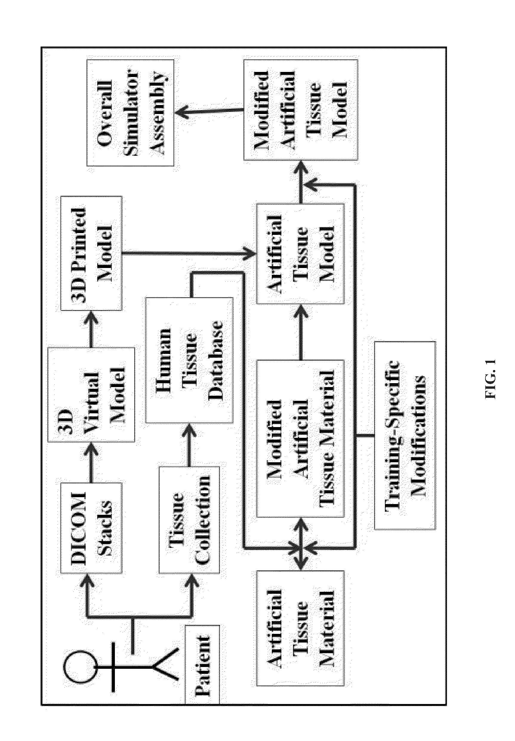
The present disclosure is about making silicone-based materials that can be used to simulate different types of tissue, such as human and animal tissue. These materials can be used for training medical or veterinary professionals and provide a realistic model for procedures that can be done on patients. The materials are made by creating a structure using a 3D printing process and then adding an artificial tissue material that has the same properties as the tissue being simulated. The artificial tissue material can also have additional indicators to help with training and procedures. The technical benefits of this patent include making a high-fidelity representation of patient tissue for training purposes and developing a tool to better prepare for procedures on patients.

Problems solved by technology

Currently, animal tissues are often used for simulation but are often anatomically different than a human patient, have different mechanical properties than human tissue with large amounts of variance between samples, are difficult to obtain and store, and have ethical issues regarding animal protection.
However, human cadaveric tissue often still has different mechanical properties than live human tissue, is typically expensive, and is difficult to obtain and store in sufficient quantities for medical training.
Cadaveric tissue also lacks the constitutive properties of fresh or live human tissue.
Neither animal tissues nor cadaveric tissues meet the fidelity needs for enhanced training, and in some cases their deficiencies can lead to negative training transfer.

Method used


Image

  • Simulated, representative high-fidelity organosilicate tissue models
  • Simulated, representative high-fidelity organosilicate tissue models
  • Simulated, representative high-fidelity organosilicate tissue models

Examples


example 1

Renal Artery Simulator

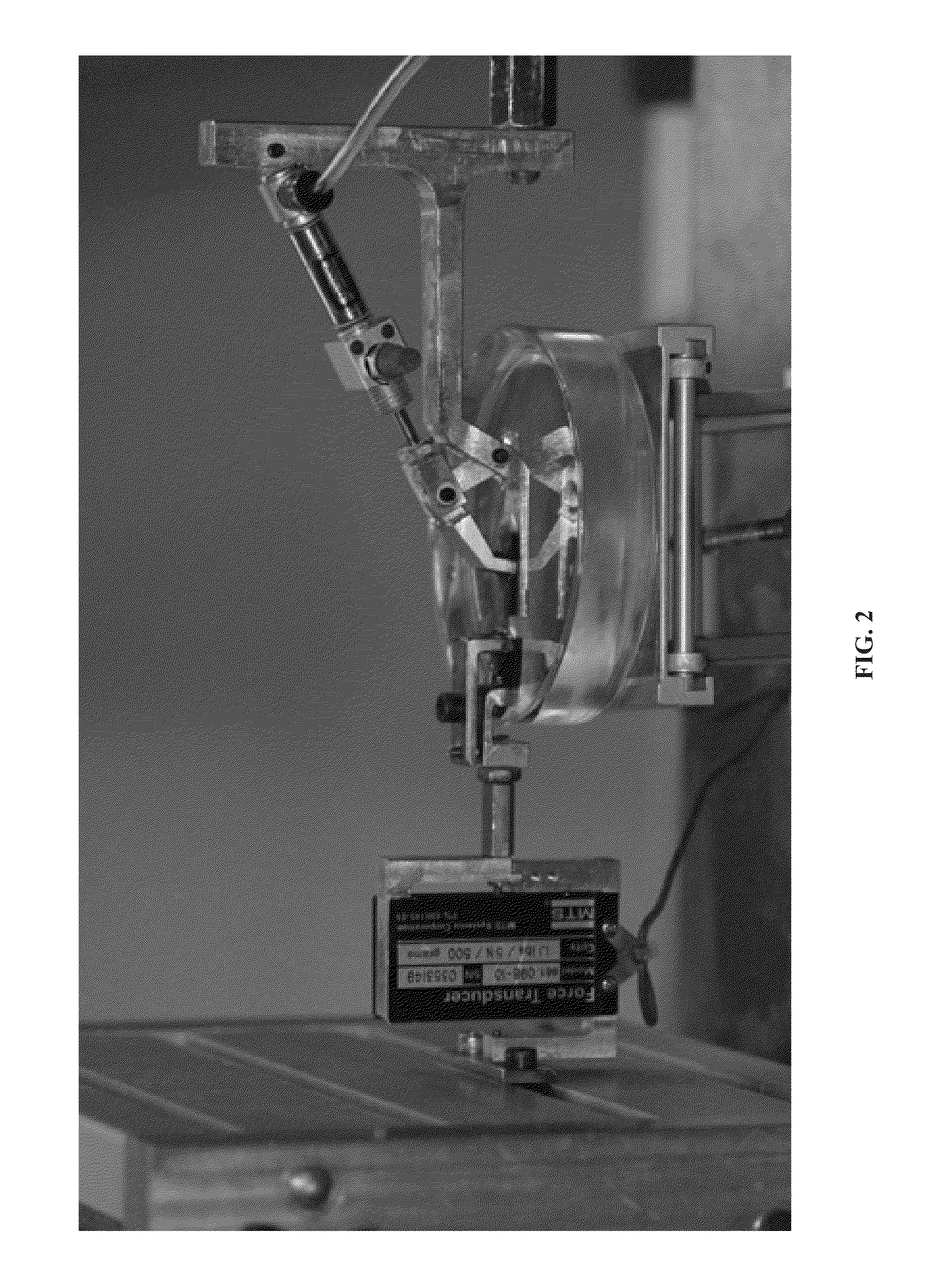
[0093]Using an organo silicate base material, the successful creation of an artificial tissue training model has been created for a human renal artery (FIG. 7) in order to meet the specifications of the American Urological Association for laparoscopic and robotic clip applying (Syverson, et al., 2011). The simulator tissue was color mapped to mimic human renal artery and filled with artificial blood to a mean arterial pressure (MAP) of 80±2 mmHG. Solid black pigment lines and dotted black pigment lines were added for training purposes to indicate areas for clipping and cutting respectively. The model was fitted into a mechanical apparatus to mimic a beating motion.

example 2

Kidney Simulator

[0094]A kidney simulator (FIG. 8) was also developed using the techniques described above. The simulator utilizes renal tissue properties, e.g., from a human tissue database, accurate human anatomical modeling (stereolithographic prototyping) and color mapping to create realistic internal features such as the endoluminal ureter and the calyceal kidney collecting system. The model can be used in combination with artificial kidney stones and fluid to simulate procedures such as a ureteroscopy (FIG. 9), retrograde pyelography, ureteral stent placement, nephro-lithotomy, laser and extracorporeal lithotripsy and kidney stone extraction.

[0095]In conclusion, the overall method for developing artificial tissue simulators for training purposes provides accurate anatomical modeling and matching of tissue properties. The materials and fabrication techniques are cost-effective and allow for the integration of indicators to properly evaluate trainee skill acquisition. The resulti...


PUM


Abstract

A method of making a tissue model comprises determining one or more material properties of a tissue, wherein the one or more material properties include at least one of mechanical properties, electroconductive properties, optical properties, thermoconductive properties, chemical properties, and anisotropic properties, creating an anatomical structure of the tissue, selecting an artificial tissue material having one or more material properties that substantially correspond to the one or more material properties of the tissue, and coupling the artificial tissue material to the anatomical structure.

Description

CROSS-REFERENCE TO RELATED APPLICATIONS[0001]The subject matter of this application is related to Reihsen et al., U.S. Provisional Patent Application Ser. No. 61 / 541,547, entitled “SIMULATED, REPRESENTATIVE HIGH-FIDELITY ORGANOSILICATE TISSUE MODELS,” filed on Sep. 30, 2011, to Reihsen et al., U.S. Provisional Patent Application Ser. No. 61 / 589,463, entitled “SIMULATED, REPRESENTATIVE HIGH-FIDELITY ORGANOSILICATE TISSUE MODELS,” filed on Jan. 23, 2012, and to Poniatowski et al., U.S. Provisional Patent Application Ser. No. 61 / 642,117, entitled “METHOD FOR ANALYZING SURGICAL TECHNIQUE USING ASSESMENT MARKERS AND IMAGE ANALYSIS,” filed on May 3, 2012, which are each incorporated by reference herein in their entirety.TECHNICAL FIELD[0002]The present disclosure is directed to models used for simulation of tissues, such as tissue models useful for providing training of medical procedures for health care providers, and in particular to human tissue models constructed with a base of organo...

Claims


Application Information

Patent Timeline
IPC IPC(8): G06G7/60
CPCG09B23/28G09B23/30G06F19/3437B33Y50/00B33Y80/00G16H50/50
Inventor REIHSEN, TROY E.SWEET, ROBERT M.BURKE, DANIEL M.
Owner RGT UNIV OF MINNESOTA
Learn More