
Testing system for determining hypoxia induced cellular damage

a testing system and hypoxia-induced technology, applied in the field of testing system for assessing cellular damage, can solve the problems of inability to provide central laboratory, large investment cost for one-time setup, and inability to receive test results in some situations

Inactive Publication Date: 2013-02-28
CALMARK SWEDEN AB
View PDF7 Cites 33 Cited by
  • Summary
  • Abstract
  • Description
  • Claims
  • Application Information

AI Technical Summary

Benefits of technology

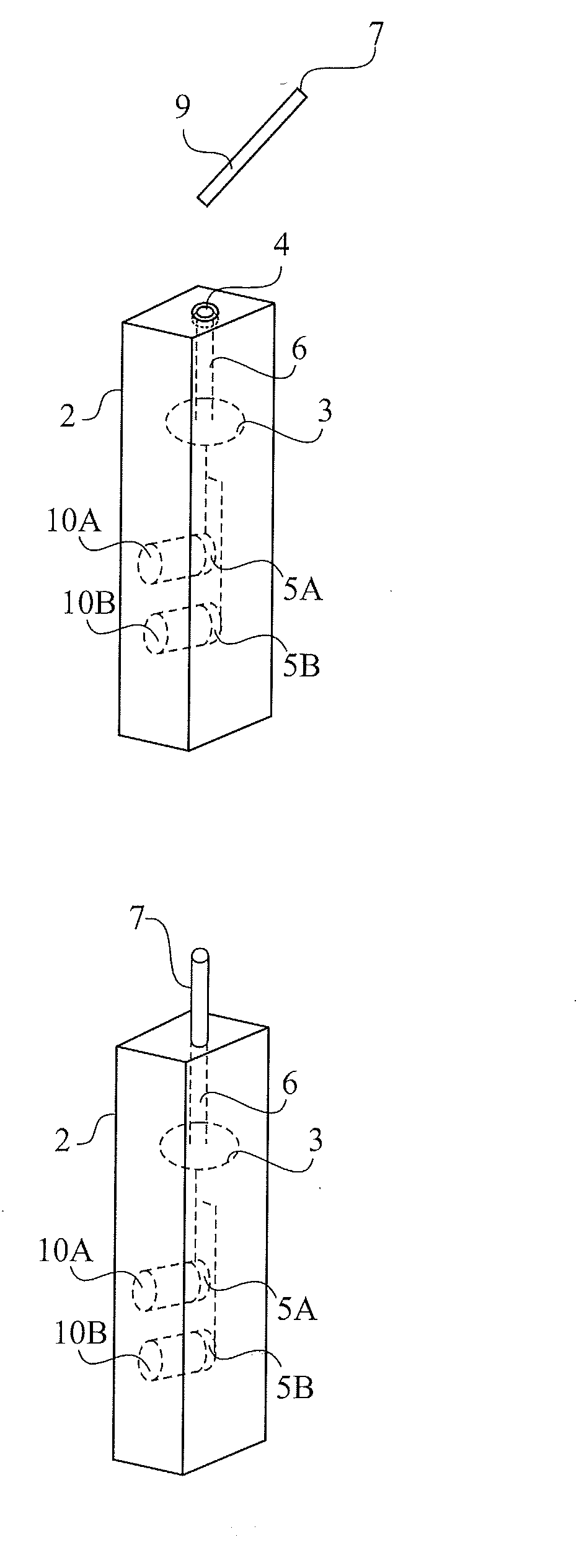
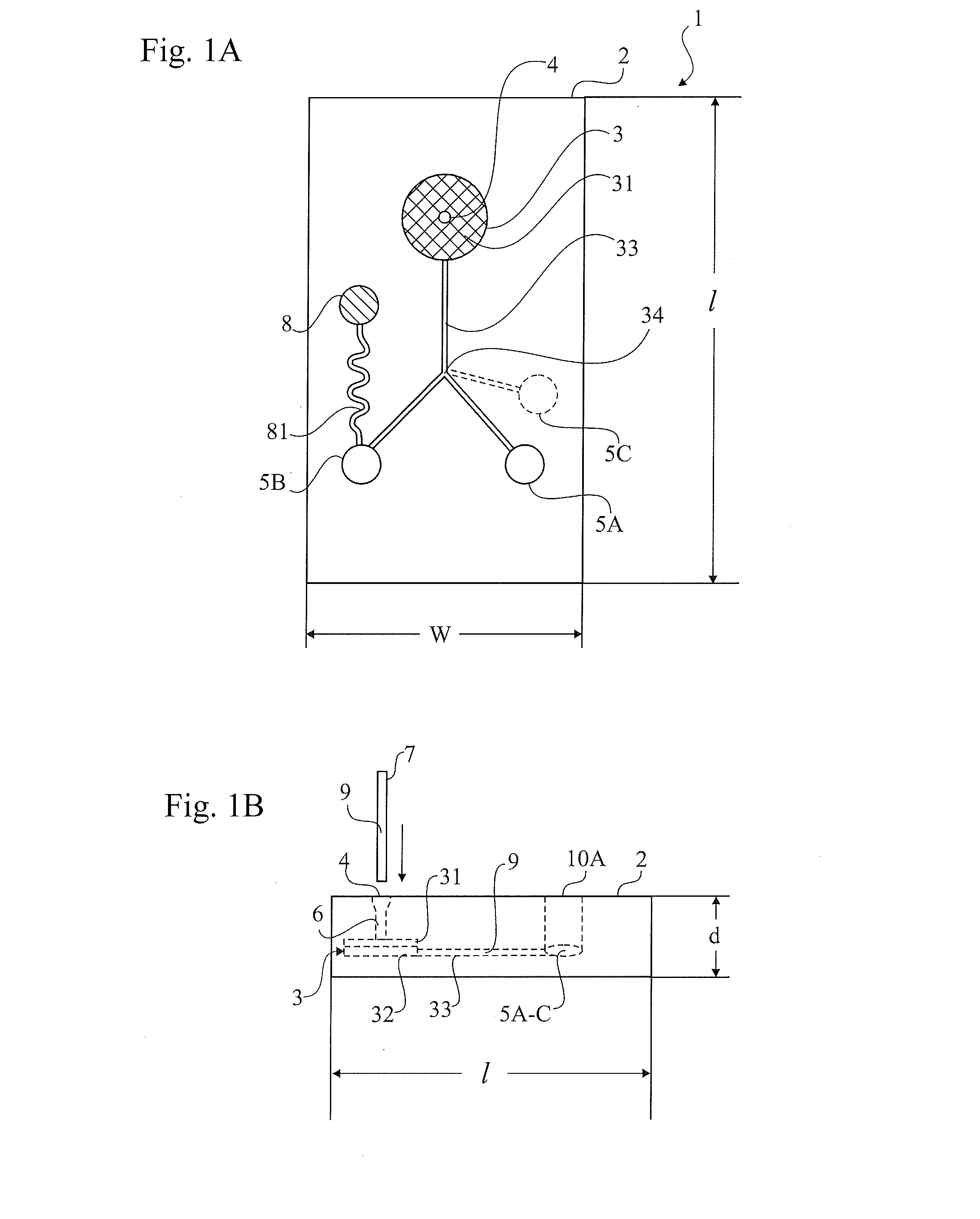
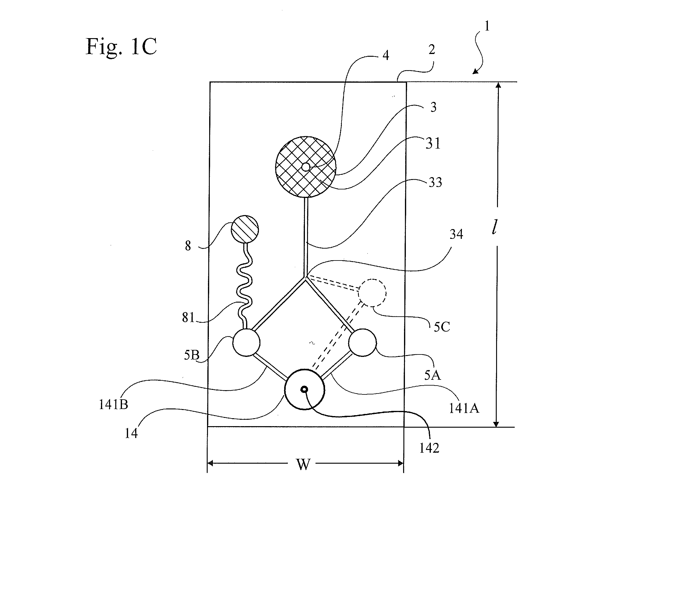
The present invention aims to provide an improved way of detecting hypoxia-induced cellular damage in a quick, user-friendly and readily available manner. It provides a small and independent test that uses a microliter-volume blood sample and visually analyzes it for selected biomarkers using a disposable device with wet chemical means. This testing system is easy to use, interpret and may be distributed as small stand-alone disposable units to medical practitioners for immediate treatment of patients. The testing system includes a wet chemical reaction designed to release the chemical means and accelerate the reaction between the reagent and any markers in the sample.

Problems solved by technology

A general problem associated with today's methods for measuring biomarkers is that they often require access to a central laboratory having the possibility of measuring marker of interest, meaning the time to receive test results in some situations becomes undesirably long.
In many places a central laboratory is not even available, and set-up of one would demand large investment costs.
Such equipments are expensive and commonly request a certain competence of the operator both to manage and interpret results of a reading.
Looking at a global perspective many countries lack a properly functioning and advanced medical treatment system, and high technology solutions may not be applicable due to lack of economical resources, or simply because of lack of physicians or health care providers who are able to perform such tests.
Even in case of a developed medical care system there are situations where long lead time and / or complicated test apparatuses are not desirable, particularly if time is crucial and a mere indication of a medical status is enough for proceeding with adequate treatment of a patient.

Method used


Image

  • Testing system for determining hypoxia induced cellular damage
  • Testing system for determining hypoxia induced cellular damage
  • Testing system for determining hypoxia induced cellular damage

Examples


example 1

[0098]Tetrazolium salts, nitro blue tetrazolium (NBT), 2-p-iodophenyl-3-p-nitrophenyl-5-phenyl tetrazolium chloride (INT) and 3-(4,5-dimethylthiazol-2-yl)-5-(3-carboxymethoxyphenyl)-2-(4-sulfophenyl)-2H-tetrazolium (MTS) were dissolved separately in dimethyl sulphoxide producing 10 mM stock solutions. The mediators phenazine methosulphate (PMS) and 1-methoxy-5-methylphenazinium methylsulfate (mPMS) were dissolved separately in water producing 1 mM stock solutions. Stock solution of NAD was prepared in buffer. Sodium lactate was dissolved in water, and pH was adjusted to about 9 with 1 M tris.

[0099]Control sera (2.2 and 4.7 μkatal / l respectively) and blood sample from co-worker were used.

[0100]Blood samples were collected in Li-heparin tubes with separator (Vacuette, Greiner) and potassium-EDTA tubes (Vacuette, Greiner). The tubes were centrifuged for 15 minutes at 1500×g and plasma was transferred into Eppendorf tubes.

[0101]Enzyme assay was performed using conventional spectrophotom...

example 2

[0104]Assays were performed using an ELISA plate reader from Emax Molecular Devices, using 96-well plates in addition to visual inspection. The bottom of the 96-well pates is used as an optical surface for measurement and each well can contain up to 400 μl liquid. Absorbance will vary depending on solution depth in wells. Plates used in this experiment were from NUNC (high binding capacity).

[0105]Tetrazolium salts, nitro blue tetrazolium (NBT), 2-p-iodophenyl-3-p-nitrophenyl-5-phenyl tetrazolium chloride (INT) and 3-(4,5-dimethylthiazol-2-yl)-5-(3-carboxymethoxyphenyl)-2-(4-sulfophenyl)-2H-tetrazolium (MTS) were dissolved separately in dimethyl sulphoxide producing 10 mM stock solutions. The mediators phenazine methosulphate (PMS) and 1-methoxy-5-methylphenazinium methylsulfate (mPMS) were dissolved separately in water producing 1 mM stock solutions. Stock solutions of NAD and NADH were prepared in buffer. Sodium lactate was dissolved in water, and pH was adjusted to about 9 with 1 ...

example 3

[0115]The following example 3 relates to wet reagent composition for assessing presence of LDH in a plasma sample.

[0116]Tetrazolium salt, nitro blue tetrazolium (NBT), was dissolved in dimethyl sulphoxide producing 10 mM stock solution. The mediator 1-methoxy-5-methylphenazinium methylsulfate (mPMS) was dissolved separately in water producing 1 mM stock solutions. Stock solution of NAD+ was prepared in buffer. Sodium lactate was dissolved in water. N-methyl-D-glucamine was dissolved in water (1M) and pH adjusted to 10 with HCl.

[0117]Control sera (2.2 and 4.7 μkatal / l respectively) and blood sample from co-worker were used.

[0118]Blood samples were collected in Li-heparin tubes with separator (Vacuette, Greiner) and potassium-EDTA tubes (Vacuette, Greiner). The tubes were centrifuged for 15 minutes at 1500×g and plasma was transferred into Eppendorf tubes.

[0119]Reaction mixture: equal volumes of tetrazolium salt stock solution, mediator stock solution, lactate and NAD+ stocks were mix...


PUM


Abstract

The present invention relates to a testing system for assessing hypoxia induced cellular damage in a mammal including human, comprising a disposable device having a sample inlet and a collection chamber separated by a separation device wherein the collection chamber is connected to at least two, a first and a second, visible detection compartments, whereof at least one is arranged with chemical means for direct visual detection, said first detection compartment being arranged to determine whether level of hemoglobin (Hb) in a sample of body fluid taken from said mammal exceeds a predetermined threshold value, and said second detection compartment being arranged to evaluate level of total amount of lactate dehydrogenase (LDH) in said sample.

Description

TECHNICAL FIELD[0001]The present invention relates to a testing system for assessing cellular damage, e.g. caused by hypoxia ischemia in a mammal including human comprising a disposable device having a sample collecting portion with a plasma separation device.BACKGROUND ART[0002]Assessment of hypoxia (oxygen deficiency) in a mammal may be done by determining total lactate dehydrogenase (LDH) within body fluid obtained from a collected sample. Measuring total amount of LDH in combination with additional prognostic markers aspartate aminotransferase (AST), alanine aminotransferase (ALT) and lactate reveal status of mammal with respect to partial or complete oxygen deficiency, information which may underlie decisions of further medical actions. Examples of medical situations where detection of hypoxia is desirable are numerous, and include perinatal and neonatal monitoring of infants, triage in emergency rooms, surgery, transplantation or other medical procedures or surgical treatments...

Claims


Application Information

Patent Timeline
IPC IPC(8): C12M1/34G01N21/78
CPCA61B5/14542A61B5/14546A61B5/1468A61B5/412A61B5/413Y10T436/201666G01N33/523G01N33/72G01N33/726G01N2333/904G01N2800/40C12Q1/32G01N33/721G01N33/54366G01N33/573
Inventor KARLSSON, MATHIASHIORT AF ORNAS, SOFIA
Owner CALMARK SWEDEN AB
Learn More