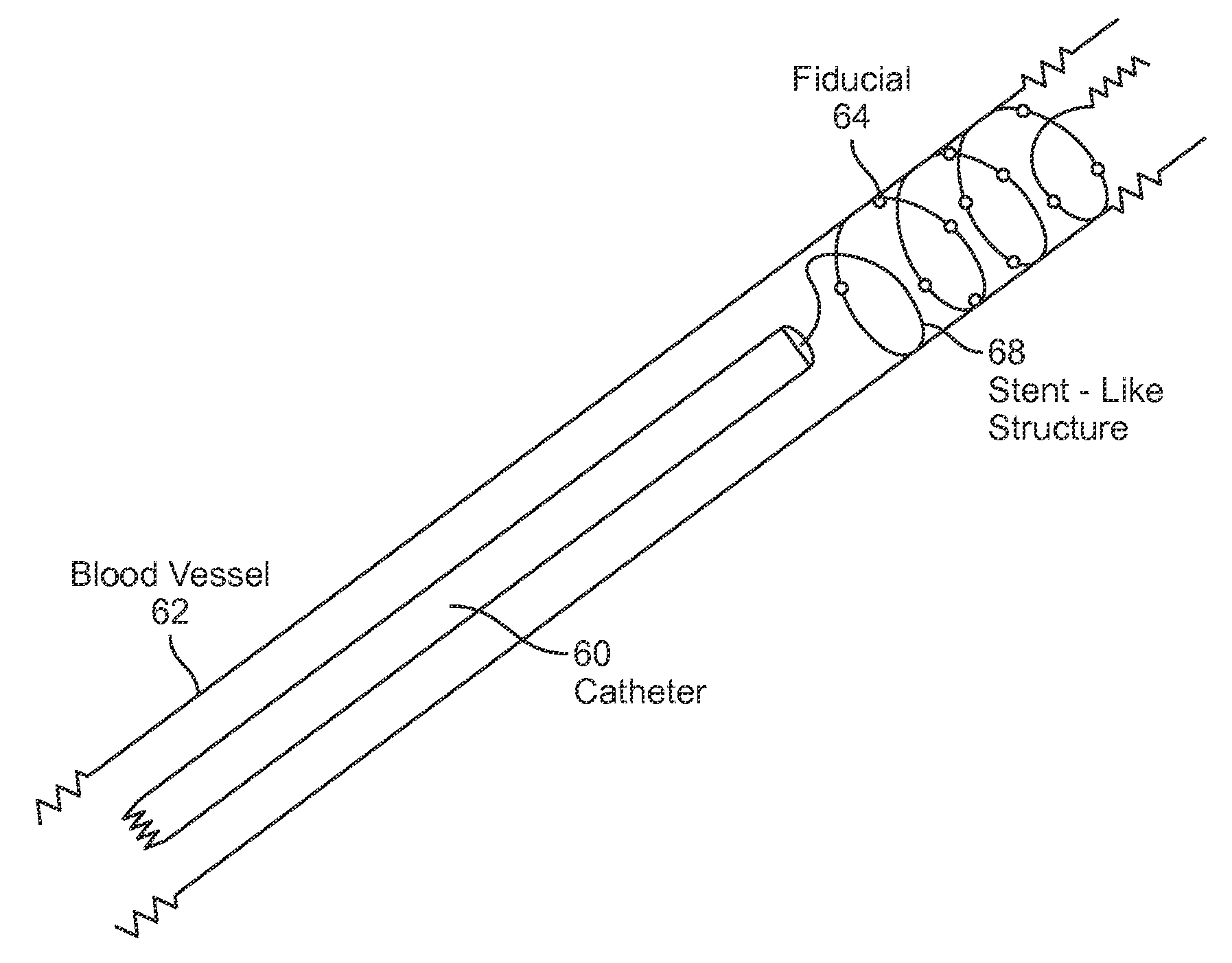
[0011]The renal nerves, owing to their position vis-à-vis the renal arteries, may correspond to moving tissue of a patient, both as a result of
respiration of the patient, the pulsation of the
aorta and
renal artery, and the
heartbeat of the patient. With respect to the latter, the temporal rise and fall of
blood pressure of the
heartbeat cycle may cause the outer
diameter of the renal arteries to expand and contract by an amount that is sufficient to move the renal nerves by an amount which may affect the treatment. Embodiments of the invention allow improved radiosurgical treatments of tissues of the renovascular
system, often enhancing the capabilities of existing radiosurgical systems for targeting what in at least some instances may be relatively rapidly moving tissues (renal nerves) so as to mitigate hypertension. Treatment of renal nerves may benefit from an implanted position surrogate for identification of the location of the
target tissue, with the surrogate optionally comprising a
fiducial marker positioned in or near the renal arteries and / or the kidneys using catheterization techniques or direct transcutaneous routes. Novel catheters and / or delivery structures having active fiducials may limit the need for X-rays (and thereby minimize collateral imaging
radiation exposure). Enhanced planning and tracking techniques may also be employed, with the radiosurgical renovascular
system treatments described herein generally being compatible with many components of existing radiosurgical treatment systems.
[0015]In many embodiments, implanting of the surrogates will comprise advancing at least one elongate flexible body through a
blood vessel. The surrogate may be coupled to tissue so that the surrogate exhibits
heartbeat- and / or respiratory-
induced movement. The implanted surrogate may comprise a non-colinear set of discrete fiducial markers so that a three-dimensional offset orientation between the surrogate and the target area can be determined from an image of the fiducial markers. In some embodiments, implanting of the surrogate may comprise screwing a
helical structure of the elongate body into a soft, contractile tissue of the heart. Implanting of the surrogate may also include expanding an expandable body with a lumen or cavity bordered by the tissue, with the expandable body optionally comprising an
inflatable balloon, a temporary
stent-like structure, or the like, which can be safely and reversibly expanded within a vessel so as to engage the surrounding tissue. In exemplary embodiments, implanting the surrogate may comprise fixing an active three-dimensional position indicator to the tissue, with the position indicator transmitting a position indicating
signal that can be used to register a location of the implanted surrogate with the planning image data. In many embodiments, the fiducial(s) will be implanted prior to acquiring planning image data. In other embodiments, an image taken after implanting the surrogate may facilitate registration. For example, when the position indicating
signal indicates an offset between the surrogate and a
position sensor (or
transmitter) disposed outside the body, the position indicating
signal can be calibrated using post-
implant image data that encompasses the
position sensor. In one exemplary embodiment, the image data used for calibrating the position indicating signal comprises post-planning calibration image data, and a calibration position sensing signal is generated while a
catheter tip engages a heart tissue. A positional relationship between the sensor and the body is maintained during acquisition of the calibration image data and the generation of the position sensing signal. More generally, the position indicator typically comprises a sensor or
signal generator used within
ultrasound or electromagnetic position indicating systems. The target region can be treated by directing the planned radiation using a position indicating signal from the position indicator between intermittent tracking
verification images. Hence, position surrogates employing such active fiducial systems may limit the need for imaging X-rays (and thereby minimize collateral imaging
radiation exposure).
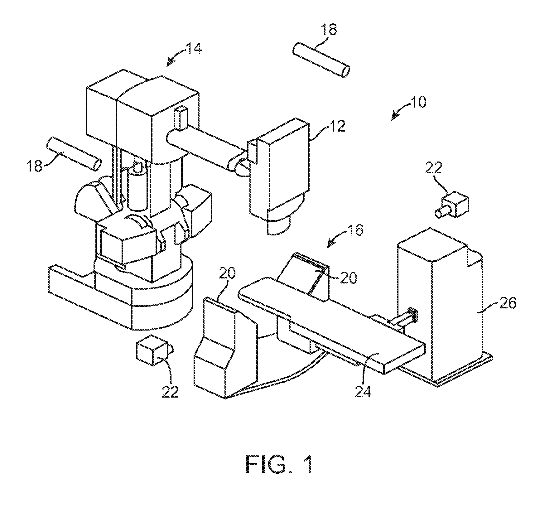
[0027]According to another aspect of the present invention, there is a radiosurgical system for treating a patient body with a
renal artery and hypertension. The system comprises an
image capture device for acquiring three dimensional planning image data from the
renal artery and / or a location
proximate the renal
artery, a radiation source for transmitting a plurality of beams of
ionizing radiation from outside the body, and a processor system configured to direct the ionizing radiation beams toward a target region of the renal
artery and / or a target region at the location proximate the renal
artery such that the radiation beams remodel the target region and the hypertension is mitigated.