
Methods for Promoting the Revascularization and Reenervation of CNS Lesions

a technology of cns and reenervation, applied in the direction of peptide/protein ingredients, prosthesis, peptide sources, etc., can solve the problems of limited success of neural transplantation for cerebral stroke repair, no effective treatment for cerebral stroke in clinical settings, and patients who survive stroke usually remain severely disabled and require constant care for the rest of their lives. , to achieve the effect of reducing the inhibition of axonal regeneration

Inactive Publication Date: 2012-11-08
MUSC FOUND FOR RES DEV +1
View PDF0 Cites 22 Cited by
  • Summary
  • Abstract
  • Description
  • Claims
  • Application Information

AI Technical Summary

Benefits of technology

The present invention relates to methods of promoting revascularization and reenervation of a CNS lesion by delivering a hydrogel containing a neural stem cell recruiting factor and / or a neural stem cell mobilizing factor to the lesion. The hydrogel can also contain other factors such as mitogens for neural stem cell proliferation / expansion, or factors that promote the differentiation of neural stem cells. The methods can be used to treat CNS lesions and prevent scar tissue growth. The technical effects include promoting the regeneration of CNS tissue and improving the recovery of neural stem cells in the lesion.

Problems solved by technology

Nonetheless, the surviving stroke patients usually remain severely disabled and require constant care for the rest of their lives.
Despite tremendous effort in thrombolysis and neuroprotection, no effective treatment is available for cerebral stroke in clinical settings.
This is largely due to the inability of current treatments to repopulate the stroke lesion cavity with functional neurons and glial cells, which dynamically participate in cell-cell signaling and provide sustained trophic support that is critical for decreased neural degeneration and sustained functional recovery.
Despite its efficacy in providing sustained functional recovery in other types of central nervous system (CNS) injuries, neural transplantation for cerebral stroke repair has had limited success, due to poor donor cell survival and functionality at the infarct site (Savitz et al., NeuroRx 1:406 (2004)).
The presence of the scar tissue not only contributes to regenerative failure, but also to the poor survival and functionality of transplanted cells, and poses a diffusive barrier that hinders the effective delivery of nutrients, oxygen, and pharmacological agents into the lesion cavity.
Secondary injury leads to delayed necrosis and apoptosis resulting in further neuronal loss.
The results from both these trials, as well as many subsequent studies focusing on the different treatment strategies, have shown no benefit in secondary injury prevention.

Method used


Image

  • Methods for Promoting the Revascularization and Reenervation of CNS Lesions
  • Methods for Promoting the Revascularization and Reenervation of CNS Lesions
  • Methods for Promoting the Revascularization and Reenervation of CNS Lesions

Examples


example 1

Composition of Synthetic Hydrogels

[0138]FIGS. 1A-C show human embryonic stem cell derived neurospheres cultured in hydrogels comprising different ratios of 4-Arm PEG and short peptide sequence (CDPVCC GTARPGYIGSRGTARCCAC). While all of the hydrogels supported growth of the cells, a PEG:peptide ratio of 25:75 produced the best results.

example 2

Sustained Release of Biologically Active Molecules grom in-situ Crosslinkable Hydrogels

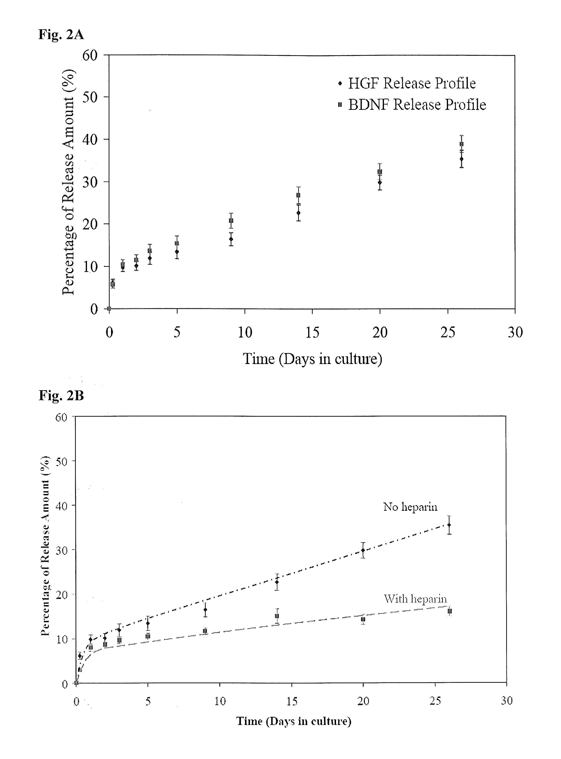
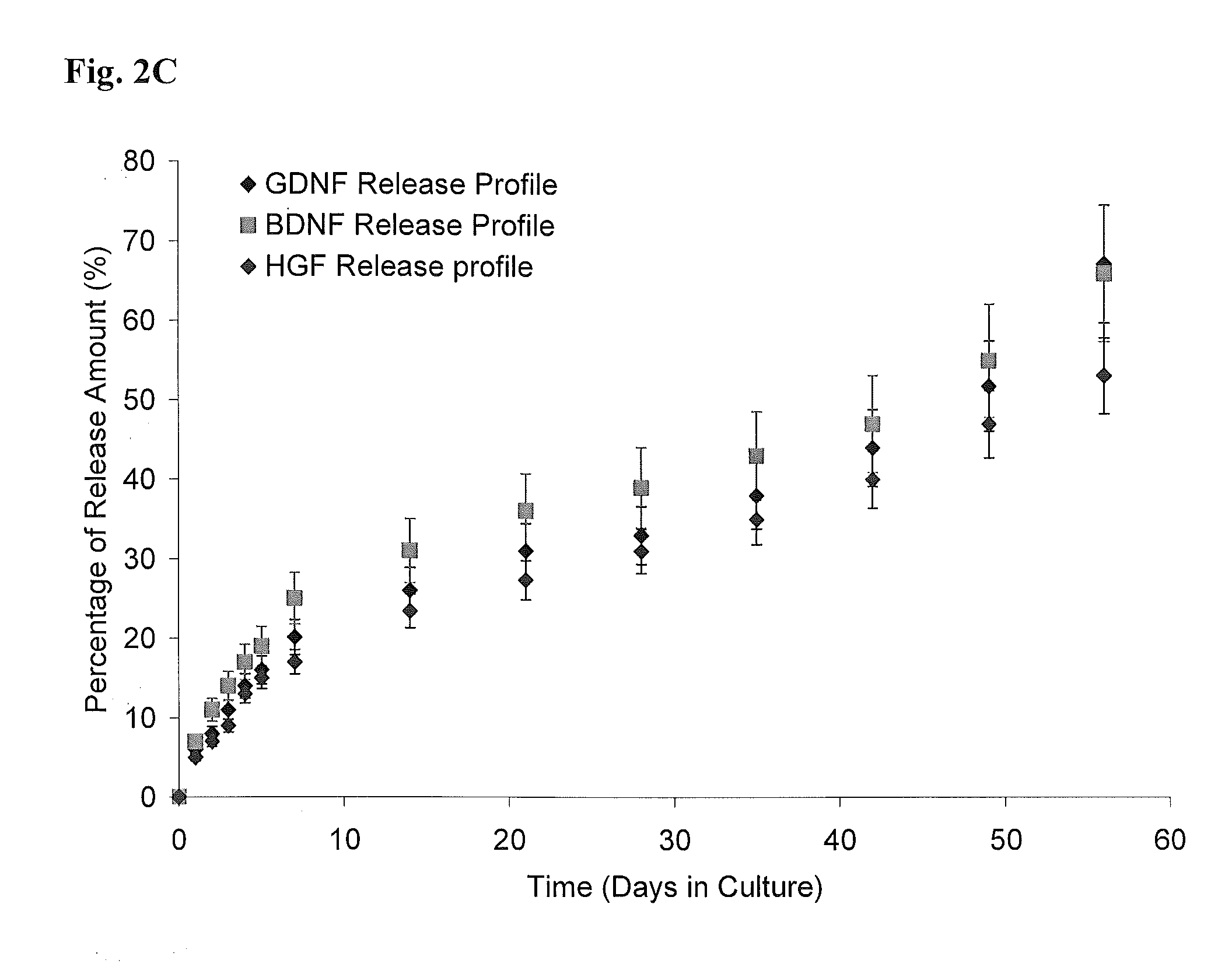
[0139]FIGS. 2A-C show sustained release of biologically active molecules from an ECM-based hydrogel. (A) Cumulative in vitro HGF and BDNF release from an ECM-based hydrogel comprising hyaluronic acid and collagen. After 26 days, approximately 35-40% of each growth factor was released from each hydrogel. (B) Cumulative in vitro HGF release from ECM-based hydrogels comprising hyaluronic acid and collagen (circles), or hyaluronic acid, collagen and heparin (squares). Addition of heparin in HA-collagen hydrogel doubles the release duration of HGF from the hydrogels. The hydrogel provides sustained release of biologically active growth factor in vitro, with release sustained for 3-6 months. This is a dramatic increase in time of availability compared to the short half-life of free growth factors in vivo. (C) Cumulative in vitro GDNF, BDNF and HGF release from the synthetic hydrogel. After 1 and 2 month...

example 3

Attracting Stem Cells in Vitro and in Vivo

[0140]FIGS. 3A-B show recruitment of stein cells to in-situ crosslinkable hydrogels containing hepatocyte growth factor (HGF). Neural stem cells (5×10³ in 200 μl culture media) were added to the upper compartment of a transwell. The lower compartment was filled with 400 μl of culture medium and an in-situ crosslinkable hydrogel as control (A), or an in-situ crosslinkable hydrogel containing 80 ng / ml solubilized HGF (B). Hydrogels were harvested following an 8-hour incubation period and stained. Sustained and localized release of HGF from the hydrogel (B) is able to induce neural stem cell migration and recruitment into the hydrogel.

[0141]FIGS. 4A-D show recruitment of endogenous stein cells to ECM-based hydrogels containing hepatocyte growth factor (HGF). ECM-based hydrogels loaded with control (A) or HGF (B) were implanted into the subcutaneous space on the back of mice. Hydrogels were harvested 1 week after implantation and samples of each...


PUM

PropertyMeasurementUnit
concentrationsaaaaaaaaaa
concentrationaaaaaaaaaa

Abstract

The present invention provides methods of promoting the revascularization and / or reenervation of central nervous system lesions using an in-situ crosslinkable hydrogel. The present invention also provides methods of treating a spinal cord injury by topically delivering to the spinal cord injury site a vehicle comprising a neurotrophic factor and / or anti-inflammatory agent. Also provided are methods of treating a spinal cord injury by topically administering or delivering a hydrogel to the injury site.

Description

STATEMENT OF GOVERNMENT SUPPORT[0001]Aspects of this invention were supported by government funding under Grant Nos. R01 NS050243 and P20RR021949 from the National Institutes of Health and Grant Nos. SC090380 and PT073600 from the Department of Defense. The U.S. Government has certain rights in this invention.FIELD OF THE INVENTION[0002]The present invention relates to methods of treating central nervous system lesions, including promoting revascularization and / or reenervation. The present invention also relates to treating spinal cord injury, including reducing inhibition of axonal regeneration and / or decreasing secondary injury at a spinal cord injury site.BACKGROUND OF THE INVENTION[0003]Brain stroke ranks as the third leading cause of death and disability in most developed countries (Wolfe et al., J. Neurol. Neurosurg. Psychiatry 72:211 (2002), and is the second most common cause of death worldwide (Murray et al., Lancet 349:1269 (1997)). About ⅙ of all human beings will suffer ...

Claims


Application Information

Patent Timeline
Patent Type & Authority Applications(United States)
IPC IPC(8): A61K38/18A61P29/00A61K9/127B82Y5/00
CPCA61K38/1825A61K38/1833A61K38/1841A61L27/52A61L27/54A61L2300/412A61L27/26A61K38/41A61K45/06B82Y5/00A61K38/185A61L2430/38A61K2300/00C08L89/06C08L5/08A61P29/00
Inventor XING, JINXUEJUN, WENKRISHNA, VIBHORZHANG, NING
Owner MUSC FOUND FOR RES DEV
Learn More