
Method for improving blood flow in bone head

a bone head and blood flow technology, applied in the field of bone head blood flow improvement, can solve the problems of bone head brittleness, reduced blood flow in the femoral head, deformation of the joint surface, etc., and achieves the effect of improving blood flow, reducing invasion, and improving blood flow

Inactive Publication Date: 2012-09-13
TERUMO KK
View PDF11 Cites 1 Cited by
  • Summary
  • Abstract
  • Description
  • Claims
  • Application Information

AI Technical Summary

Benefits of technology

[0008]It is an object of the present invention to provide a method for improving the blood flow in the bone head, especially the femoral head with a minimum invasion on the patient.
[0009]It is another object of the present invention to provide a method for improving the blood flow in the femoral head, thereby treating the ischemic disease that causes the osteonecrosis in the femoral head.
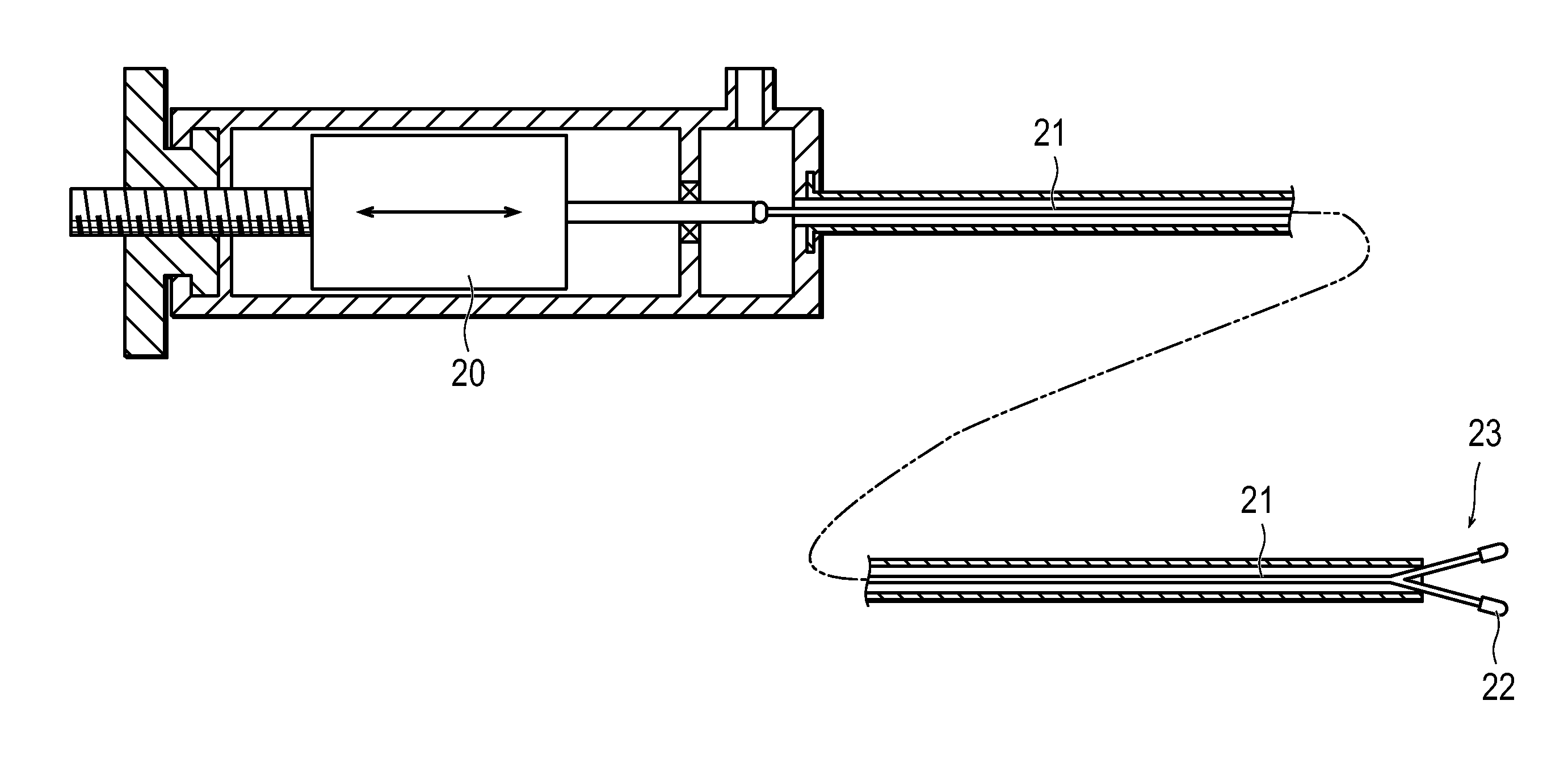
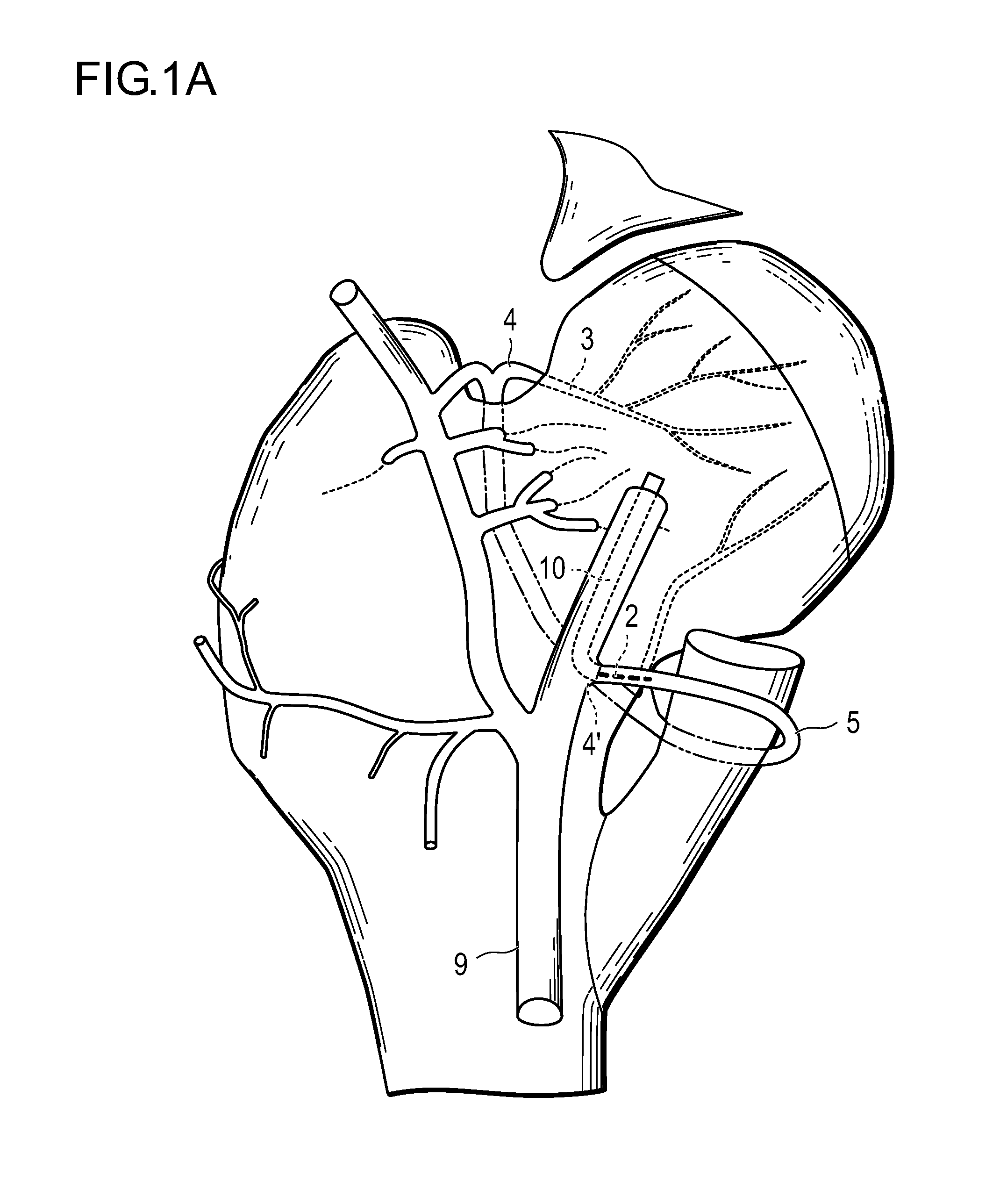
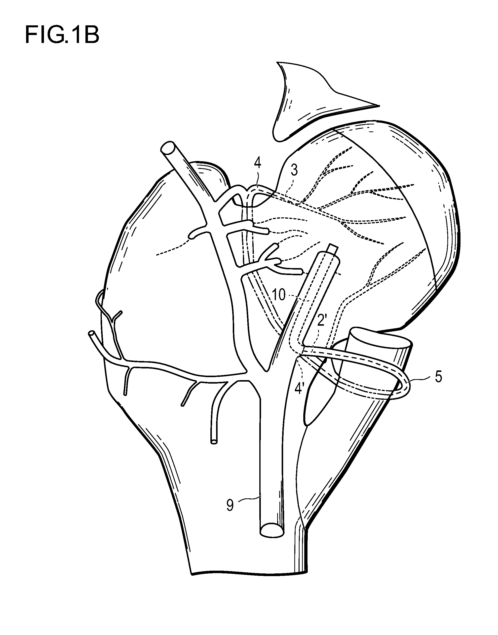
[0010]According to its one embodiment, the present invention provides a method for improving the blood flow in the bone head, the method including the steps of extending a long tubular body, which has a cutting tool at its foreend, close to the entrance of the retinaculum artery and performing drilling on the bone head by using the cutting tool.
[0011]The method according to the present invention improves the blood flow in the head of the bone, particularly the head of the femur, with a minimum burden on the patient.

Problems solved by technology

Particularly, the osteonecrosis that occurs in the femoral head (the upper end of the femur held in the hip joint at the root of the leg) leading bony tissues to death, thereby causing depression or deformation to the joint surface.
It reduces the blood flow in the femoral head and makes the bony tissues brittle due to necrosis.
The progression of this symptom leads to the crushing of the femoral head and the osteoarthrosis of the hip joint, which brings about pains and gait disturbance, thereby greatly deteriorating the quality of life (QOL).
The cause of the osteonecrosis of the femoral head has not yet been completely cleared up; however, it appears without any cause or it appears relatively frequently in those who drink alcohol in large amounts or who experience systemic administration of steroid in large amounts.
However, the conservative treatment is intended to relieve the symptom but is not intended to completely cure the osteonecrosis of the femoral head.
In addition, because the conservative treatment is not fully expected to prevent the progress of crushing, the operative treatment is performed for conservation of the femoral head in the case where crushing is likely to progress.
Unfortunately, the operative treatment mentioned above has to be performed under general anesthesia and needs the patient to be hospitalized for 5 to 7 days, depending on his age and conditions, followed by 6 weeks to 3 months for complete recovery.
Moreover, the patient requires rehabilitation for a long period of time until he becomes capable of daily life without help.
As mentioned above, the osteonecrosis in the femoral head, once it appears, is hard to cure by conservative treatment and needs time and cost for surgical treatment.
Thus, it causes a large damage to medical and social economy.
Under these circumstances, early diagnosis and early treatment are desirable but there has been no effective method for treatment to prevent the progression of the disease.

Method used


Image

  • Method for improving blood flow in bone head
  • Method for improving blood flow in bone head
  • Method for improving blood flow in bone head

Examples


example 1

[0075]A patient suffering from femoral head necrosis undergoes X-ray or MRI examination to ascertain the range of necrosis. At least 24 hours before insertion of a catheter, the patient is orally given clopidogrel (as an antiplatelet agent) (300 mg) once a day on the first day of administration. If antithrombotic treatment is necessary, the patient is orally given it (75 mg for maintenance dose) once a day at the same timing as above.

[0076]The patient has a sheath placed in the femoral artery of the other one of the patient's leg having a lesion, with the help of a catheter introducer kit (Radifocus Introducer, made by Terumo Corporation). Through this sheath is inserted a guide wire, 0.035 inches in diameter (Radifocus Guide Wire M, made by Terumo Corporation). The guide wire is advanced under X-ray radioscopy until its foreend reaches the inside femoral circumflex artery through the deep artery of thigh from the femoral artery. Along the guide wire is inserted a guiding catheter 4...

example 2

[0078]A patient suffering from femoral head necrosis undergoes X-ray or MRI examination to ascertain the range of necrosis. At least 24 hours before insertion of a catheter, the patient is orally given clopidogrel (as an antiplatelet agent) (300 mg) once a day on the first day of administration. If antithrombotic treatment is necessary, the patient is orally given it (75 mg for maintenance dose) once a day at the same timing as above.

[0079]The patient has a sheath placed in the femoral artery of the other one of the patient's leg having a lesion, with the help of a catheter introducer kit (Radifocus Introducer, made by Terumo Corporation). Through this sheath is inserted a guide wire, 0.035 inches in diameter (Radifocus Guide Wire M, made by Terumo Corporation). The guide wire is advanced under X-ray radioscopy to the femoral artery where there exists the lesion and then inserted to the point which is slightly beyond the branch point of the deep artery of thigh and the outside femor...

example 3

[0081]The same procedure as in Example 2 is repeated except that the cutting catheter is advanced to the point which is beyond the epyphysis line of the femoral head and 3 mm inside the foreend of the bone head, with slow injection from a syringe inserted into the catheter hub at the end of the rotablator. The syringe contains 0.5 mL of alprostadil as a vasodilator, and the rate of injection is 50 μL / min (or 250 ng / min of alprostadil).


PUM


Abstract

A method for improving the blood flow in the bone head, the method including the steps of extending a long tubular body, which has a cutting tool at its foreend, close to the entrance of the retinaculum artery and performing drilling on the bone head by using the cutting tool. This method makes it possible to improve the blood flow in the bone head with a minimum of burden on the patient.

Description

BACKGROUND OF THE INVENTION[0001]1. Field of the Invention[0002]The present invention relates to a method for improving the blood flow in the bone head. More particularly, the present invention relates to a method for improving the blood flow in the bone head (such as the head of the femur or humerus) suffering from osteonecrosis and also to a method for healing ischemic diseases leading to osteonecrosis.[0003]2. Description of the Related Art[0004]Osteonecrosis is one of the ischemic diseases that occurs in the head of the femur, the head of the humerus, or the condyle of the femur. Particularly, the osteonecrosis that occurs in the femoral head (the upper end of the femur held in the hip joint at the root of the leg) leading bony tissues to death, thereby causing depression or deformation to the joint surface. It reduces the blood flow in the femoral head and makes the bony tissues brittle due to necrosis. The progression of this symptom leads to the crushing of the femoral head a...

Claims


Application Information

Patent Timeline
IPC IPC(8): A61B17/16
CPCA61B17/1615A61B17/1617A61B2017/564A61B17/1675A61B17/1684A61B17/1668
Inventor NAKAGAWA, YUJIKINOSHITA, YASUSHITADA, YUICHIHATA, SUGURU
Owner TERUMO KK
Learn More