Method for producing pluripotent stem cells
a technology of stem cells and cells, applied in the field of stem cell production methods, can solve the problems that human cells have not yet been able to achieve the technique of producing pluripotent stem cells
- Summary
- Abstract
- Description
- Claims
- Application Information
AI Technical Summary
Benefits of technology
Problems solved by technology
Method used
Image
Examples
example 1
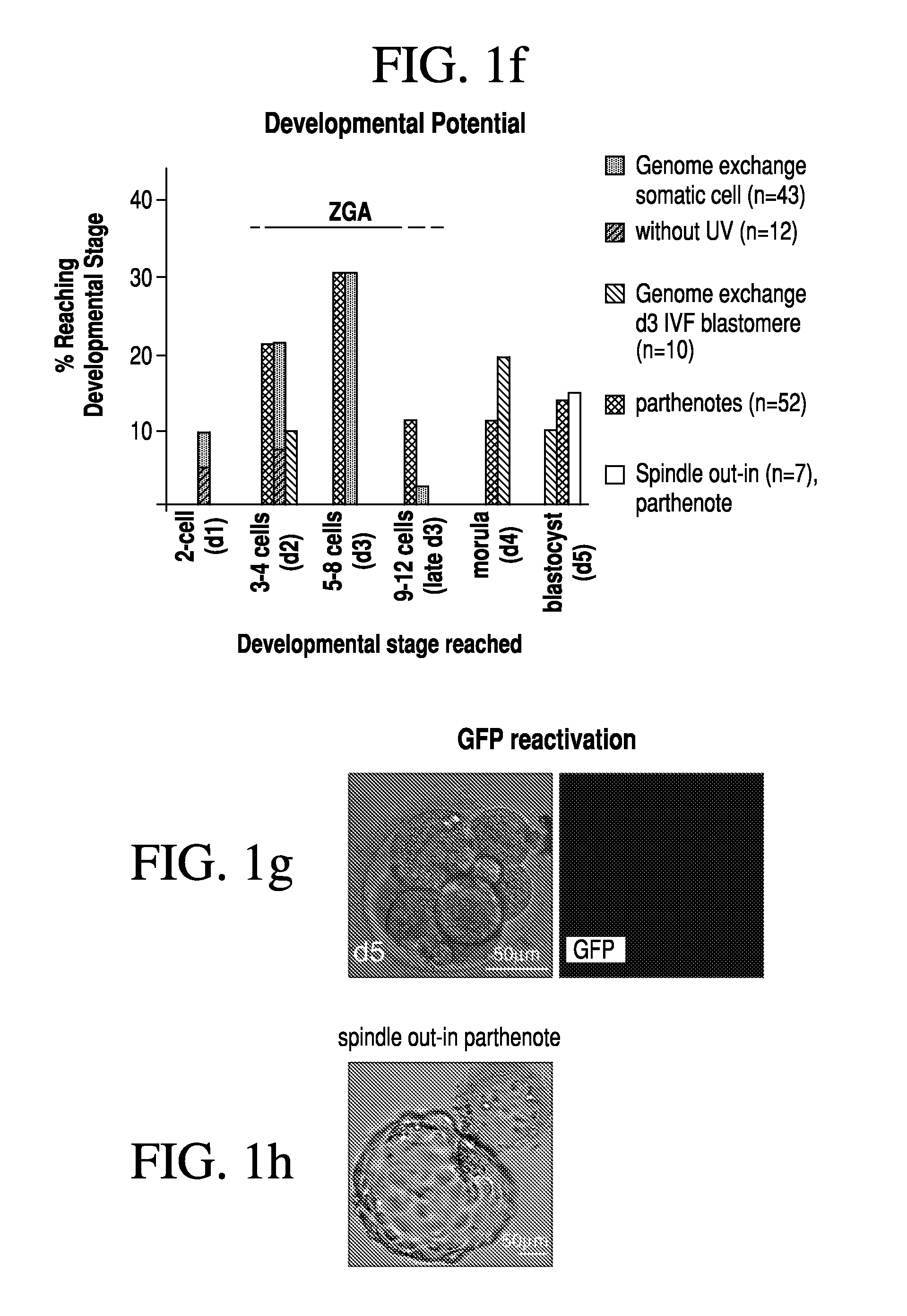
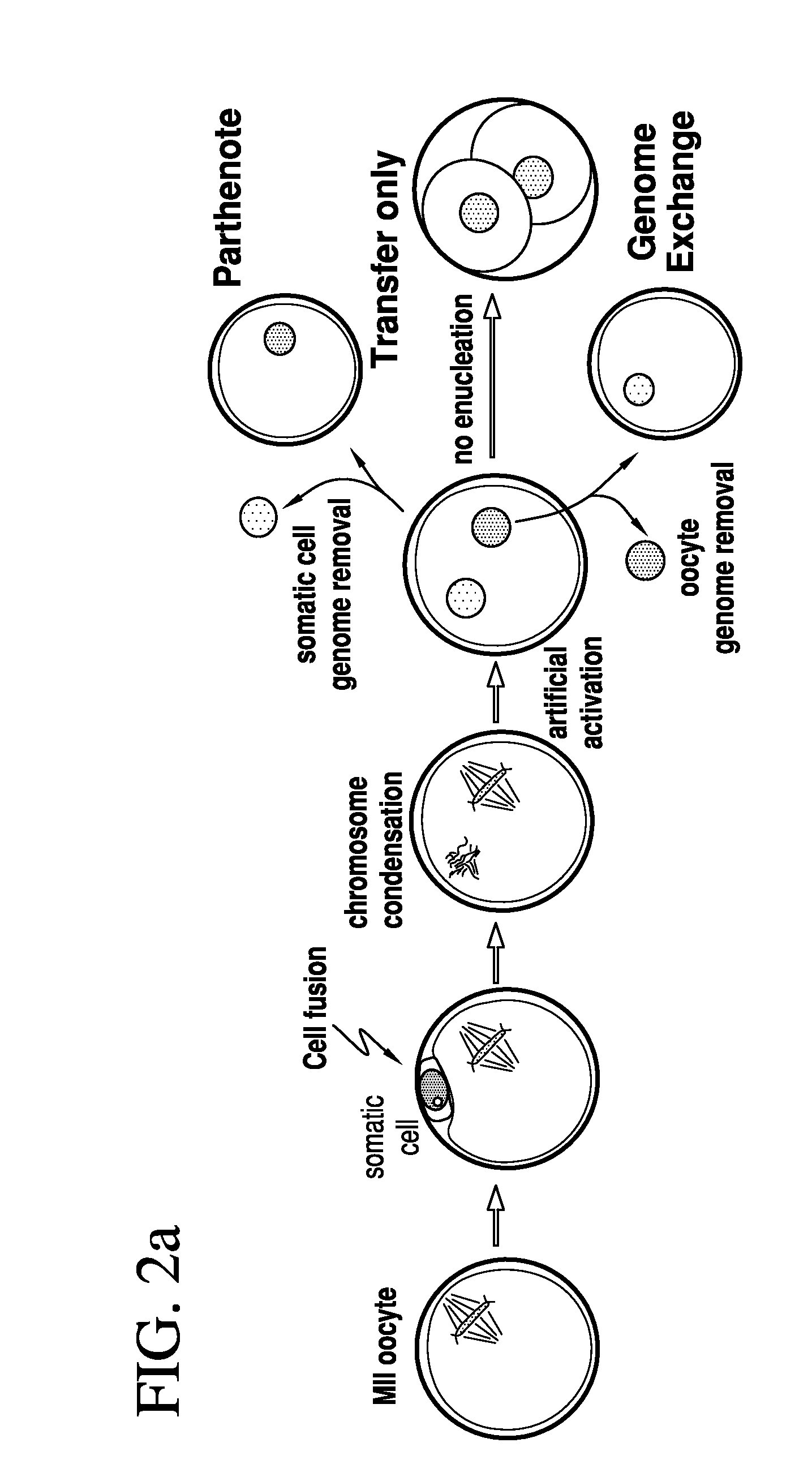
[0079]Two methods were developed for nuclear transfer. For all methods, the somatic cells were allowed to grow to confluence to induce growth arrest, optionally followed by 1 day of incubation in medium with low serum content (0.5% fetal bovine serum). Prior to transfer, somatic cells were lifted from the dish using trypsin (Invitrogen), pelletted by centrifugation (1000 g for 4 min), resuspended in fibroblast medium (Dulbecco's Modified Eagle Medium [“DMEM”] plus 10% FBS, Invitrogen) and kept on ice until transfer. The oocytes were retrieved from healthy young donors (22-33 years of age). The somatic cell was introduced into the oocyte by an 20 μs 1.3 kV / cm electrical pulse in cell fusion medium 0.26M mannitol, 0.1 mM MgSO4, 0.05% BSA, 0.5 mM 4-(2-hydroxyethyl)-1-piperazineethanesulfonic acid (“HEPES”). The electrical pulse was delivered with an Electro Cell Fusion Generator, LF201, (Nepagene). The same type of pulse was used to transfer an oocyte genome or a blasto...
example 2
Artificial Activation
[0081]At 3-4 hours post nuclear transfer, when chromosome condensation of the somatic cell has occurred, the oocyte was activated in 5 μM ionomycin followed by 4-6 hours in 2 mM 6-dimethylaminopurine (“6-DMAP”). The inonomycin was diluted in GMOPSplus containing HSA (Vitrolife). Embryos were then allowed to develop in embryo culture media at 5% oxygen, 6% CO2 and 89% N2, in. Global media supplemented with 10% plasmanate, in a Cook benchtop incubator. Embryo culture could also be done under ambient oxygen concentrations, and using different commercial embryo culture media, or a different incubator.
example 3
Genome Removal
[0082]Three methods have been developed for the removal of a genome from the oocyte.
[0083]In one such method, the oocyte genome was identified at the metaphase II stage with spindle birefringence using the Oosight™ system (CRi) and then removed in the presence of 5-10 μg / ml cytochalasinB using a fire-polished pipette (as the one in FIG. 6c). The pipette was prepared in our laboratory using a Sutter needle puller and a micro-forge (Sutter instruments). In approx. a third of the nuclear transfer experiments, genome removal was done in this manner. In another method, the location of the oocyte genome was identified at the metaphase II stage by staining in 2 μg / ml of Hoechst 33342 and minimal UV illumination and then removed in the presence of 5-10 μg / ml cytochalasinB. Minimal UV illumination is the lowest illumination that still allows the visualization of the chromosomes by eye in a dark room and after the eyes are adapted to darkness. Minimal UV illumination is achieved...
PUM
| Property | Measurement | Unit |
|---|---|---|
| Time | aaaaa | aaaaa |
| Time | aaaaa | aaaaa |
| Mass | aaaaa | aaaaa |
Abstract
Description
Claims
Application Information
 - R&D
- Intellectual Property
- Life Sciences
- Materials
- Tech Scout
- Unparalleled Data Quality
- Higher Quality Content
- 60% Fewer Hallucinations
Browse by: Latest US Patents, China's latest patents, Technical Efficacy Thesaurus, Application Domain, Technology Topic, Popular Technical Reports.
© 2025 PatSnap. All rights reserved.Legal|Privacy policy|Modern Slavery Act Transparency Statement|Sitemap|About US| Contact US: help@patsnap.com


