
Method and apparatus for determining heart rate variability using wavelet transformation

a wavelet transformation and heart rate technology, applied in the field of advanced signal processing methods, can solve the problems of lack of appropriate surgical attention, time-consuming and expensive computational devices, and inability to detect p and t waves

Inactive Publication Date: 2012-05-17
J FITNESS LLC +1
View PDF3 Cites 470 Cited by
  • Summary
  • Abstract
  • Description
  • Claims
  • Application Information

AI Technical Summary

Benefits of technology

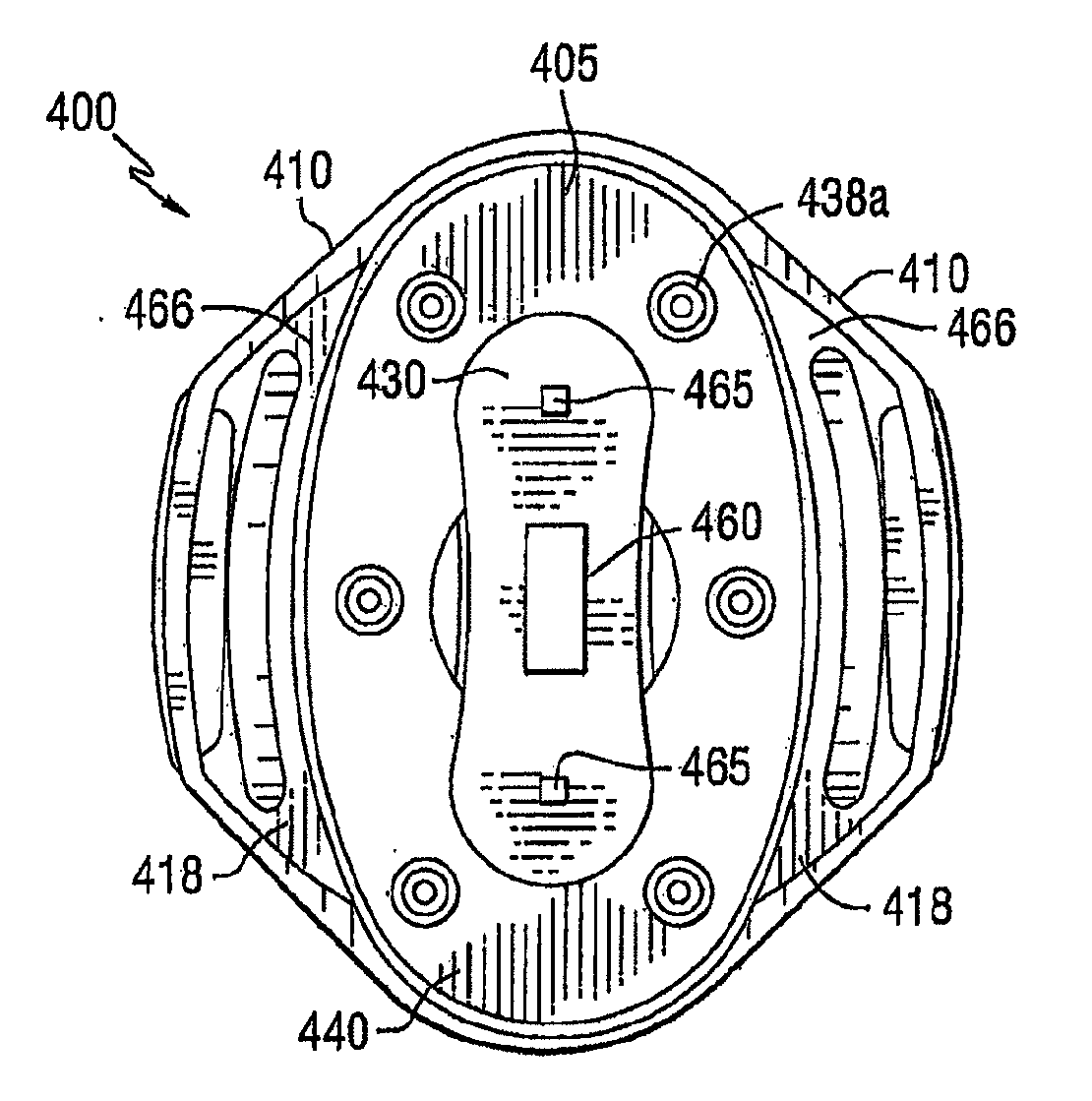
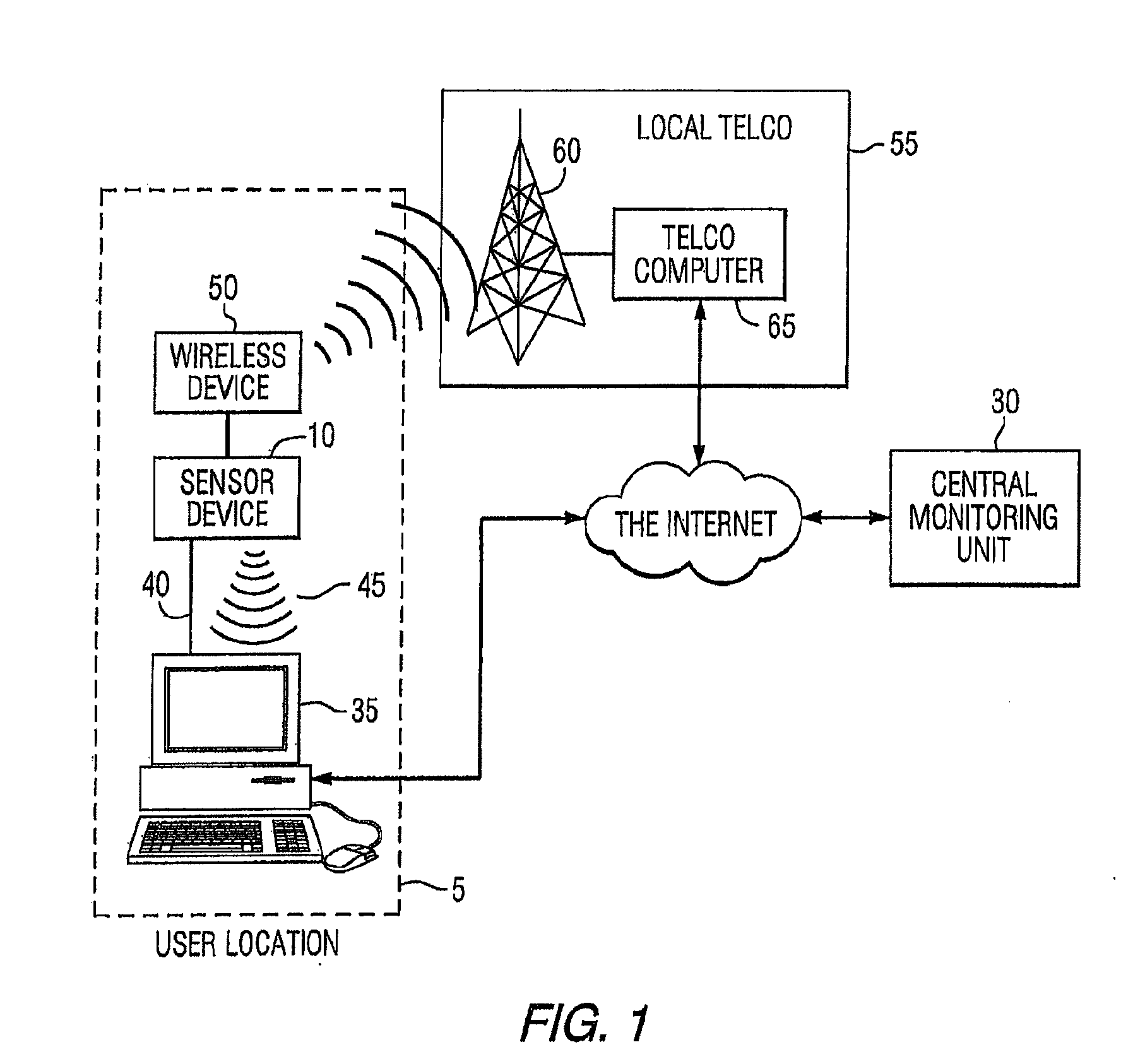
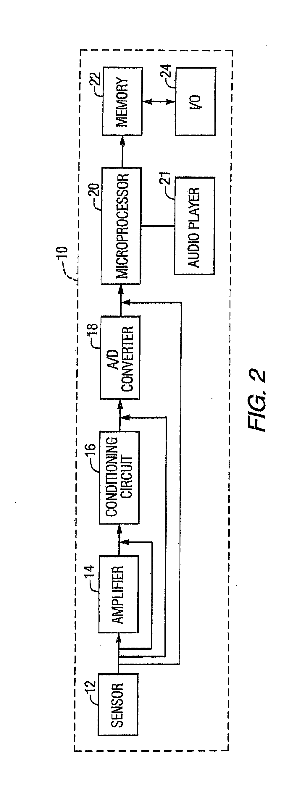
The present invention relates to an apparatus for detecting and displaying the mammalian ECG waveform. The apparatus collects data from sensors that are worn on the individual's body and uses mathematical operations to determine the presence of heart-related parameters. It can help care workers estimate the extent of blood volume loss, distinguish between physical activity and exercise, and predict the presence of cardiovascular disease. The apparatus is designed for long-term wear and can be used in conjunction with a software platform to monitor critical heart-related measures. It is easy to enter and track physical information and can detect and analyze ECG waveforms in real-time. The apparatus is comfortable and convenient for individuals to wear and can be used in various activities of daily life.

Problems solved by technology

Literature shows enormous methods for ECG components detection, however, it is sparse for P and T wave detection.
Many prior studies have focused only on detection of the QRS-complex because P and T waves are sparse and harder to isolate from the signal.
Unfortunately, mathematical modelings of ECG segment and threshold methods are sensitive to noise and baseline drift artifacts exist in the signal which can unexpectedly affect the detection.
Although WT methods currently exist in the literature and are transparent to noise and baseline drift artifacts, they are time consuming and require more powerful computational devices.
This is problematic, given the utility of WT in ECG analysis for detection of its components more accurately and effectively in terms of both speed and memory requirements.
Acute traumatic shock resulting in tissue injury and hemorrhage remains the primary cause of death on the battlefield, and is also a leading cause of death in civilian trauma.
Both these rapid deaths and many complications associated with the injury result from a lack of appropriate surgical attention and limited evacuation facilities in the field.
However, the treatment of battlefield injuries is more challenging and fatalities within the first hour of wounding are highly dependent on battlefield conditions.
These conditions include the availability of medical personal and their skill and experience; limitations of medical equipment; and delay in transit to the nearest medical facility [.
Field treatment of injuries has thus been a priority research issue.
However, the potential risks and benefits of early fluid resuscitation have been studied previously, with the conclusion that careful evaluation is required before changes are made to the established treatment methods for trauma patents.
However, PSD estimation methods are unsuitable for analyzing series whose characteristics change rapidly [.
Also, spectral analysis of the residual ECG is sometimes difficult due to a low signal-to-noise ratio (SNR).
These difficulties are worsened when time-frequency analysis with a short duration time window is used.
Rickards et al found that the measure of high frequency to low frequency (HF / LF) alone may not be enough to differentiate between LBNP and physical activity even though the measure has the potential different between normal and disease subject.
Therefore, the ability to differentiate heart rate changes from blood loss due to wounding and heart rate changes due to activity have been inconclusive.
Hemorrhagic shock (HS) can be a lethal consequence of injury sustained on the battlefield as well as in civilian life.
Monitoring the health status of combatants using easily obtained signals such as heart rate remains a challenge.
Unfortunately, traditional HRV analysis appears now to be unable to distinguish between central volume loss and exercise.
This is problematic given the desire to use changes in heart rate to detect the presence of acute volume loss due to hemorrhage.
In addition, little has been done to examine other physiologic signals for pattern changes indicative of critical changes in physiology in response to injury and treatment.
Lastly, almost nothing has been done in the area of using the techniques of machine learning (ML) to enhance the predictive power of signal analyses as they relate to critical illness and injury and other clinical entities.

Method used


Image

  • Method and apparatus for determining heart rate variability using wavelet transformation
  • Method and apparatus for determining heart rate variability using wavelet transformation
  • Method and apparatus for determining heart rate variability using wavelet transformation

Examples


example 1

A. Data Specification

[0292]In this study, 53 cases belonging to the Lower Body Negative Pressure (LBNP) database were used. One purpose of (LBNP) chambers is to simulate the transition from micro-gravity to Earth-gravity. Physiological tests are conducted to assess stresses upon the cardiovascular system during these simulations. In general, the internal negative air pressure of LBNP chambers is controlled with a proportional control system using only air-pressure as input. In each test, the case experienced multi-stages air pressure where in each stage, the level of negative air pressure is increased for 5 minutes, and the ECG signal is sampled at 500 Hz. Table 6 shows the LBNP protocol for each stage.

TABLE 6LBNP PROTOCOL WHEN MEASURING THE BCG SIGNALLBNP protocolStageTime0mmHgBaseline5 Min−15mmHgStage 15 Min−30mmHgStage 25 Min−45mmHgStage 35 Min−60mmHgStage 45 Min−70mmHgStage 55 Min−80mmHgStage 65 Min−90mmHgStage 75 Min−100mmHgRecovery5 Min

B. Preprocessing

[0293]As FIG. 40 shows, t...

example 2

A. Dataset and Experiment Procedure

[0322]The dataset comprises fifty-nine subjects, including forty-six LBNP subjects and thirteen exercise subjects. LBNP testing is done using a chamber in which the subject is exposed to varying levels of negative pressure. All measures are either continuously monitored or semi-continuously monitored at regular intervals. Table 10 represents detail dataset information containing LBNP and exercise subjects as well as monitoring timeframe for LBNP protocol. Cardiovascular collapse is defined for LBNP as the stage the protocol was terminated dues to physical or physiologic signs or symptoms of distress. Collapse state for exercise was defined as the stage of exercise resulting in a matched heart rate from the same subject who also underwent LBNP study. Recovery stage indicates removing the negative pressure from the subjects. The exercise protocol comprised: 5 minutes baseline followed by 5 minute levels of exercise at gradually increasing workloads. ...

example 3

[0356]Two studies were performed:

Study 1: Comparison of LBNP vs. Exercise:

[0357]A repeated ANOVA analysis is performed with Turkey post-hoc tests to compare the HRV response over 4 stages (baseline until stages 4) of LBNP and exercise subjects.

Study 2: Classification of LBNP Stages

[0358]The general task of classification is to predict the specific LBNP stage for any given input ECG based on the study of training examples. Also, Arterial Blood Pressure and impedance signals are added to ECG for classification. The aim of using ML algorithms is to generate a simple classification function which is easy to understand. Thus, three machine learning algorithms are used: SVM, AdaBoost, and C4.5.

[0359]For each study, a total of forty-five features are extracted, thirty-six features for DWT (db 32) and DWT (db4), seven for PSD, and two features for Higuchi FD. In Study 1, ANOV A analysis is performed using the cardiovascular response over 3 stages (baseline plus 3 stages) of LBNP with 4 stag...


PUM


Abstract

The present invention relates to advanced signal processing methods including digital wavelet transformation to analyze heart-related electronic signals and extract features that can accurately identify various states of the cardiovascular system. The invention may be utilized to estimate the extent of blood volume loss, distinguish blood volume loss from physiological activities associated with exercise, and predict the presence and extent of cardiovascular disease in general.

Description

CROSS REFERENCE TO RELATED APPLICATIONS[0001]This application is a continuation-in-part of International Application No. PCT / US09 / 06234, titled “Method and Apparatus for Determining Critical Care Parameters,” filed Nov. 20, 2009. PCT / US09 / 06234 is a continuation-in-part of U.S. application Ser. No. 11 / 928,302, filed on Oct. 30, 2007, which is a continuation of U.S. application Ser. No. 10 / 940,889, filed Sep. 13, 2004, issued as U.S. Pat. No. 7,502,643. U.S. application Ser. No. 10 / 940,889 claims the benefit of U.S. Provisional Application Ser. No. 60 / 502,764, filed Sep. 12, 2003; U.S. Provisional Application Ser. No. 60 / 510,013, filed Oct. 9, 2003; and U.S. Provisional Application Ser. No. 60 / 555,280, filed Mar. 22, 2004. PCT / US09 / 06234 is also a continuation-in-part of co-pending U.S. patent application Ser. No. 10 / 940,214, filed Sep. 13, 2004, which is a continuation in part of co-pending U.S. application Ser. No. 10 / 638,588, filed Aug. 11, 2003, which is a continuation of U.S. ap...

Claims


Application Information

Patent Timeline
Patent Type & Authority Applications(United States)
IPC IPC(8): A61B5/0205A61B5/08A61B5/145A61B5/01A61B5/053A61B5/0488A61B6/00A61B5/0476A61B3/113A61B5/107A61B5/11A61B5/1468A61B5/22A61B5/021A61B5/044G16Z99/00
CPCA61B5/0022A61B5/7267A61B5/02125A61B5/024A61B5/02405A61B5/0402A61B5/0476A61B5/0488A61B5/0496A61B5/053A61B5/0816A61B5/0833A61B5/1116A61B5/1118A61B5/14532A61B5/4872A61B5/4875A61B5/7203A61B5/02055G16H40/67A61B5/398A61B5/318A61B5/389A61B5/369G16Z99/00
Inventor NAJARIAN, KAYVANANDRE, DAVIDWARD, KEVINVYAS, NISARGTELLER, ERICSTIVORIC, JOHN M.FARRINGDON, JONATHANBOEHMKE, SCOTT K.KOVACS, GREGORYGABARRO, JAMESKASABACH, CHRISTOPHERJI, SOO-YEONRAOFF, ABEL ALPELLETIER, RAYMOND
Owner J FITNESS LLC
Learn More