Lung cancer diagnosis

a lung cancer and cancer technology, applied in the field of cancer diagnosis, can solve the problems of not being able to achieve the routine screening for lung cancer or early detection of the disease, unnecessary and potentially harmful invasive procedures and/or therapeutic regimens, and not being able to achieve the effect of reducing the burden of tumors and high throughpu

Inactive Publication Date: 2012-04-26
FRED HUTCHINSON CANCER RES CENT
View PDF0 Cites 7 Cited by
  • Summary
  • Abstract
  • Description
  • Claims
  • Application Information

AI Technical Summary

Benefits of technology

[0013]Several proteomics methods are emerging as useful means for discovering autoantibody biomarkers (e.g., Hanash, Nature 422:226-32, 2003; Imafuku, Omenn and Hanash, Dis Markers 20:149-53, 2004; U.S. Pat. Nos. 6,645,465; 7,202,045; 7,387,881). The merit of a proteomic approach is that it allows identification of autoantibodies to proteins that are directly derived from cancer cells or tumors and thus may uncover antigenicity associated with proteins as they occur in tumor cells, including proteins whose antigenicities have structural bases in their post-translational modification. Previous studies using two-dimensional gels of lung tumor cell lysates and Western blotting uncovered autoantibodies in lung cancer patient sera against annexin I, PGP9.5 and 14-3-3 theta proteins (Brichory et al., Cancer Res 61:7908-12, 2001; Brichory et al., Proc Natl Acad Sci USA 98:9824-9, 2001; Pereira-Faca et al., Cancer Res 67:12000-6, 2007).
[0014]More recently, a method has been implemented that utilized liquid-based procedures to separate intact proteins in tissue and tumor cell lysates (Wang and Hanash, J Chromatogr B Analyt Technol Biomed Life Sci 787:11-8, 2003; Faca et al., J Proteome Res 6:3558-65, 2007). Several hundreds of distinct protein-containing fractions were spotted onto microarrays, interrogated using various sources of sera, and quantitatively analyzed for bound antibodies. Anti-PGP9.5 antibodies were successfully identified in sera of newly diagnosed lung cancer patients, and anti-UCHL3 antibodies were identified i...

Problems solved by technology

In its early stages, lung cancer produces no symptoms, thereby typically eluding detection until the disease has progressed to advanced stages that are associated with high mortality rates.
These approaches have not, however, been shown to reduce lung cancer mortality rates, and may generate false positive results that could lead to unnecessary and potentially harmful invasive procedures and / or therapeutic regimens.
Hence, these procedures are not amenable to routine screening for, or early detection of, lung cancer.

Method used


Image

  • Lung cancer diagnosis
  • Lung cancer diagnosis
  • Lung cancer diagnosis

Examples


example 1

Materials and Methods

Materials

[0073]Nitrocellulose-coated FAST slides were purchased from Whatman (Sanford, Me.). Alexa 647-labeled anti-human IgG and recombinant protein arrays were purchased from Invitrogen (Carlsbad, Calif.).

Serum Samples

[0074]Serum samples and controls were obtained following informed consent. Sera from newly diagnosed lung cancer patients and matched controls were collected through the Community Clinical Oncology Program at the University of Michigan. Pre-diagnostic blood samples from lung cancer patients and matched controls were randomly chosen in pairs from the CARET serum bank (Goodman et al., J Natl Cancer Inst 96:1743-50, 2004; Omenn et al., N Engl J Med 334:1150-5, 1996). The distribution of histology and time from blood draw to diagnosis for the 85 pre-diagnostic lung cancer cases are shown in Tables 1 and 2.

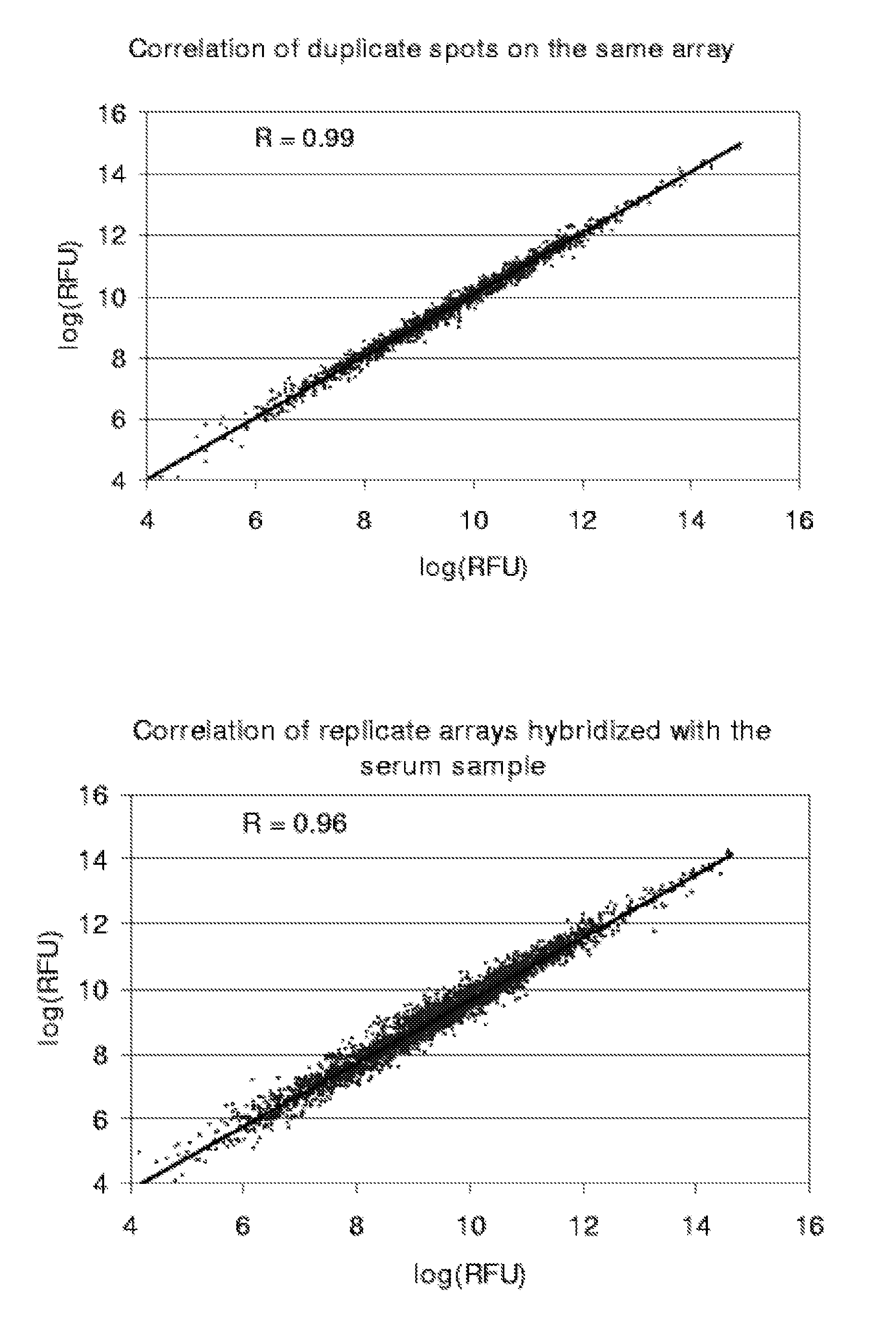
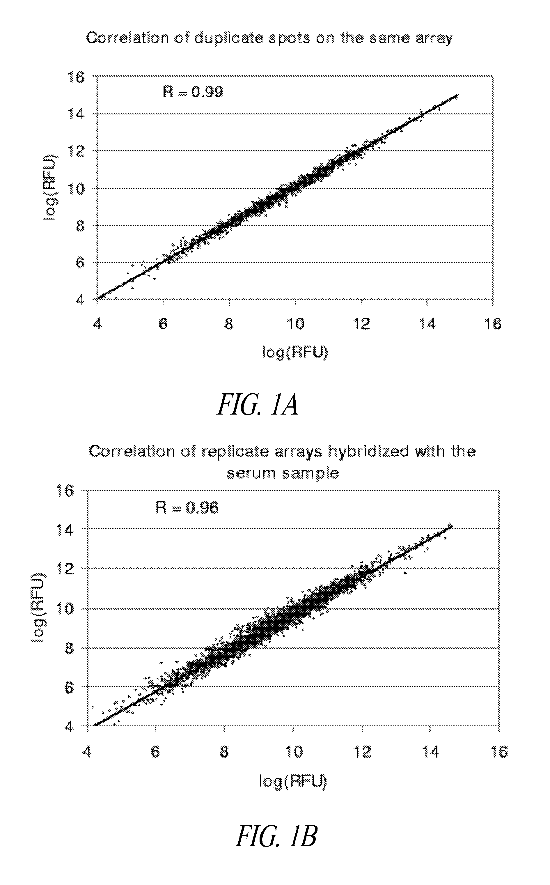
[0075]Natural Protein Microarray Production

[0076]50 mg proteins from the human lung adenocarcinoma cell line A549 lysates were first separated by a...

example 2

Results

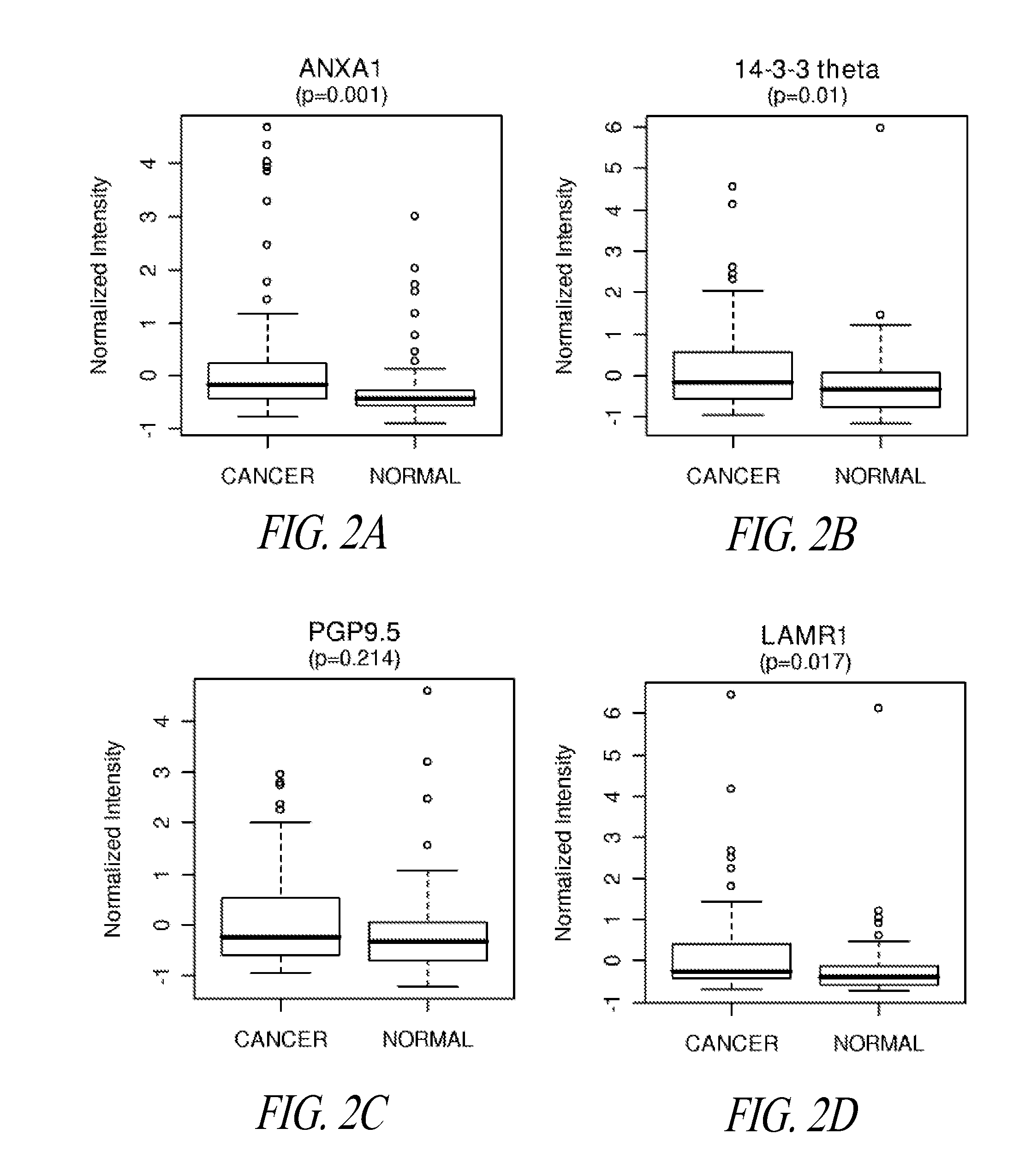
[0085]Validation Study of Autoantibodies to Annexin I, PGP9.5, and 14-3-3 Theta in Pre-Diagnostic Sera

[0086]Annexin I, PGP9.5 and 14-3-3 theta were previously identified as inducing an autoantibody response in lung cancer, based on 2D Western analysis of sera from newly diagnosed subjects with lung cancer (Brichory et al., Cancer Res 61:7908-12, 2001; Brichory et al., Proc Natl Acad Sci USA 98:9824-9, 2001; Pereira-Faca et al., Cancer Res 67:12000-6, 2007). Natural protein microarrays were developed to screen tumor-derived proteins for antigens that induce autoantibodies, based on extensive protein fractionation followed by spotting of aliquots from individual fractions (Madoz-Gurpide et al., Proteomics 1:1279-87, 2001). Natural protein containing microarrays were utilized to investigate the occurrence of autoantibodies to annexin I, PGP9.5 and 14-3-3 theta reactivity in pre-diagnostic sera. Serum specimens from the CARET cohort, which consisted of subjects at increased risk ...

example 3

Exemplary Pre-Diagnostic Lung Cancer Indicator Proteins

[0105]

ANNEXIN 1>ipi|IPI00218918|IPI00218918.5 ANNEXIN A1 (SEQ ID NO: 1):MAMVSEFLKQAWFIENEEQEYVQTVKSSKGGPGSAVSPYPTFNPSSDVAALHKAIMVKGVDEATIIDILTKRNNAQRQQIKAAYLQETGKPLDETLKKALTGHLEEVVLALLKTPAQFDADELRAAMKGLGTDEDTLIEILASRTNKEIRDINRVYREELKRDLAKDITSDTSGDFRNALLSLAKGDRSEDFGVNEDLADSDARALYEAGERRKGTDVNVFNTILTTRSYPQLRRVFQKYTKYSKHDMNKVLDLELKGDIEKCLTAIVKCATSKPAFFAEKLHQAMKGVGTRHKALIRIMVSRSEIDMNDIKAFYQKMYGISLCQAILDETKGDYEKILVALCGGN>ipi|IPI00549413|IPI00549413.2 ANNEXIN A1 (SEQ ID NO: 2):MNLILRYTFSKMAMVSEFLKQAWFIENEEQEYVQTVKSSKGGPGSAVSPYPTFNPSSDVAALHKAIMVKGVDEATIIDILTKRNNAQRQQIKAAYLQETGKPLDETLKKALTGHLEEVVLALLKTPAQFDADELRAAMKGLGTDEDTLIEILASRTNKEIRDINRVYREELKRDLAKDITSDTSGDFRNALLSLAKGDRSEDFG>ipi|IPI00643231|IPI00643231.1 ANNEXIN A1 (SEQ ID NO: 3):MAMVSEFLKQAWFIENEEQEYVQTVKSSKGGPGSAVSPYPTFNPSSDVAALHKAIMVKGVDEATIIDILTKRNNAQRQQIKAAYLQETGKPLDETLKKALTGHLEEVVLALLKTP14-3-3 THETA>ipi|IPI00018146|IPI00018146.1 14-3-3 PROTEIN THETA (SEQ ID NO: 4):MEKTELIQKAKL...


PUM


Abstract

Diagnosis of lung cancer in a subject before onset of symptoms is described herein (i.e., in a pre-diagnostic subject), by screening a biological fluid from the subject for the presence therein of autoantibodies that are specific for one or more pre-diagnostic lung cancer indicator proteins, including LAMR1, and optionally additionally or alternatively including annexin I and / or 14-3-3-theta and / or other pre-diagnostic lung cancer indicator proteins as presently disclosed, as the defined antigens. Related methods, including for monitoring immune reactivity against lung cancer indicator proteins in a lung cancer patient, typing lung cancer subjects or characterizing lung tumors, and application of the described proteomics approach for the identification of additional pre-diagnostic lung cancer indicator proteins, are also contemplated.

Description

CROSS REFERENCE TO RELATED APPLICATION[0001]This application claims the benefit of U.S. Provisional Application Ser. No. 61 / 095,269, filed Sep. 8, 2008.STATEMENT OF GOVERNMENT INTEREST[0002]This invention was made with government support under Grant No. UO1 CA084982, awarded by the National Cancer Institute / National Institutes of Health. The government has certain rights in this invention.STATEMENT REGARDING SEQUENCE LISTING[0003]The Sequence Listing associated with this application is provided in text format in lieu of a paper copy, and is hereby incorporated by reference into the specification. The name of the text file containing the Sequence Listing is 360056—404PC_SEQUENCE_LISTING.txt. The text file is 65 KB, was created on Aug. 27, 2009 and is being submitted electronically via EFS-Web to the U.S. PCT Receiving Office, concurrent with the filing of the specification.BACKGROUND[0004]1. Technical Field[0005]The presently disclosed invention embodiments relate to compositions and...

Claims


Application Information

Patent Timeline
IPC IPC(8): G01N33/53
CPCG01N33/57423
Inventor HANASH, SAMIR M.QIU, JIFACA, SANDRA
Owner FRED HUTCHINSON CANCER RES CENT
Why Eureka
Try Eureka