Visual stimulation presenting apparatus, functional magnetic resonance imaging apparatus, magnetoencephalograph apparatus, and brain function measurement method

a technology of visual stimulation and presenting apparatus, which is applied in the field of visual stimulation presenting apparatus, functional magnetic resonance imaging apparatus, magnetoencephalograph apparatus, and brain function measurement method, can solve the problems of high quality, and difficult to completely reproduce the above-described process of light source, object and reflection. , to achieve the effect of fine surface state and high degree of texture of objects

Inactive Publication Date: 2011-12-01
CANON KK
View PDF7 Cites 10 Cited by
  • Summary
  • Abstract
  • Description
  • Claims
  • Application Information

AI Technical Summary

Benefits of technology

[0017]Aspects of the present invention are directed to a visual stimulation presenting apparatus, a functional magnetic resonance imaging apparatus, a magnetoencephalograph apparatus, and a brain function measurement method, enabling, at the time of running an apparatus to perform brain function measurement of the subject by detecting change in magnetic field, with regard to selectively presenting to a subject multiple specimens for visual stimulation which are made up of actual objects, realization of precise control of presenting time and presenting position, and presenting a fine surface state of and a high degree of sense of texture of the objects.
[0023]According to the present invention, with regard to selectively presenting to a subject multiple specimens for visual stimulation which are made up of actual objects, a visual stimulation presenting apparatus, a functional magnetic resonance imaging apparatus, a magnetoencephalograph apparatus, and a brain function measurement method, can precisely control the presentation time and presenting position to realize presentation of a fine surface state of and a high degree of sense of texture of objects at the time of running an apparatus to perform brain function measurement of the subject by detecting change in magnetic field.

Problems solved by technology

This means that in the event of performing measurement by fMRI while presenting visual stimulation, electronic equipment such as normal displays or projectors cannot be installed in the shielding room, so various contrivances have been made regarding the apparatuses to provide the stimulation to be presented to the subject during measurement by fMRI so that no electromagnetic noise is generated.
However, the conventional arrangements described above have the following problems.
However, with a case of using a project which shows images by reflecting or transmitting an image off or through a display or screen using self-emitted light to present an image of an object to an observer, it is extremely difficult to completely reproduce the above-described process of light source, object, and reflection.
Particularly, it is generally difficult to represent with high quality the sense of texture, gloss, metallic sheen, and so forth, of the surface of objects using a display or projector, so the sense of reality and presence is understood to be lower as compared to a case of observing an actual object.
Accordingly, with a technique of presenting the subject with visual stimulation using a projector or the like such as with Japanese Patent Laid-Open No. 2004-160086, the process of controlling the illumination of the actual object so as to present to the subject is absent, so it is difficult to reproduce states and the like the same as with presenting the actual object.
As a result, performing measurement by fMRI while presenting the subject with such objects with delicate surface state and a high degree of sense of texture, with a sense of presence, is difficult.
However, the target images here are presented by human hand, and it is difficult with such a technique to precisely control the presentation time and presenting position of multiple visual stimulation specimens to be presented to the subject.
2489-2495, a control unit for compressed air is installed at a position around 2.5 m away from the MRI, and a piston-shaped actuator which is installed within the MRI bore and operates with compressed air is driven, so the mechanism is complicated.
Also the positional control precision has a margin of around ±5 mm, which is not necessarily satisfactory for a highly precise control technology.

Method used


Image

  • Visual stimulation presenting apparatus, functional magnetic resonance imaging apparatus, magnetoencephalograph apparatus, and brain function measurement method
  • Visual stimulation presenting apparatus, functional magnetic resonance imaging apparatus, magnetoencephalograph apparatus, and brain function measurement method
  • Visual stimulation presenting apparatus, functional magnetic resonance imaging apparatus, magnetoencephalograph apparatus, and brain function measurement method

Examples


first embodiment

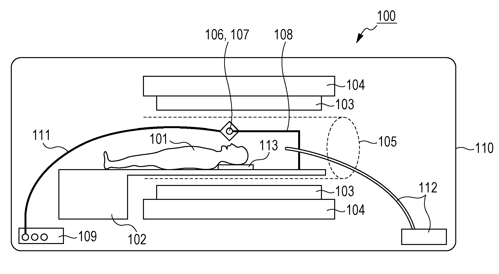
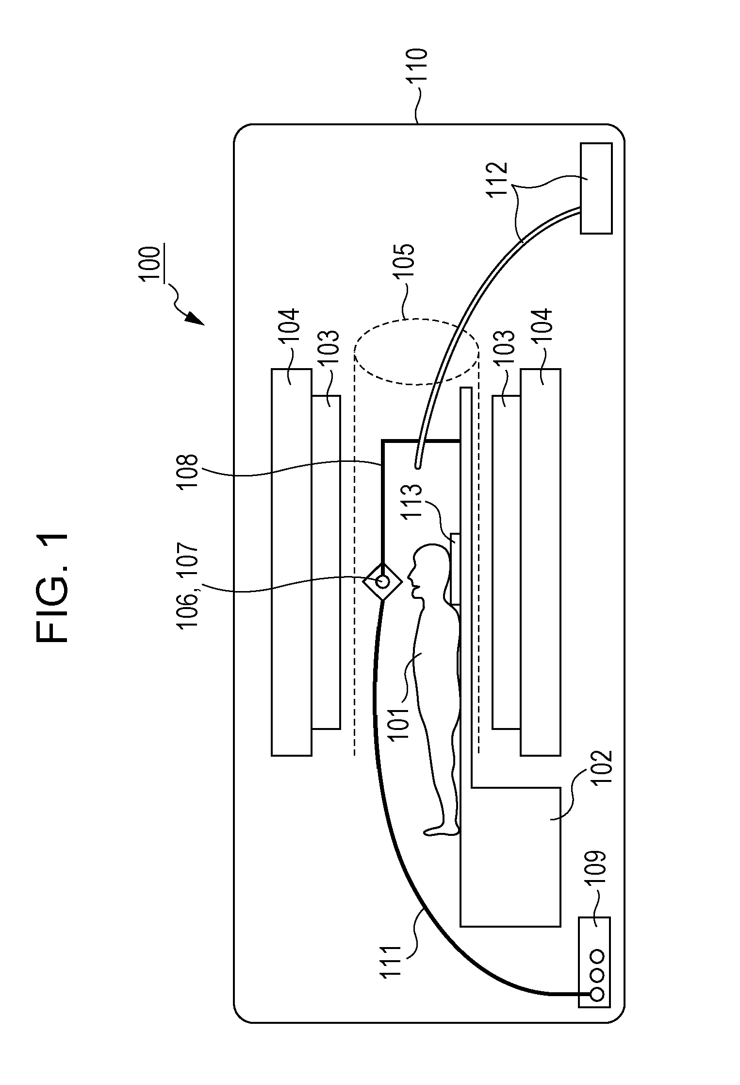
[0039]In the first embodiment, description will be made regarding an example of installing a visual stimulation presentation apparatus for presenting multiple specimens for visual stimulation that are made up of actual objects to the subject, in a functional magnetic resonance imaging (fMRI) apparatus, and performing brain function measurement. Using a functional magnetic resonance imaging (fMRI) apparatus is preferable, since active portions of the brain in manifestation of brain functions can be imaged with high sensitivity and highly fine images.

[0040]First, in the present embodiment, a configuration example in a case of installing a visual stimulation presentation apparatus for selectively presenting multiple specimens for visual stimulation in a functional magnetic resonance imaging apparatus which performs brain function measurement of a subject will be described with reference to FIG. 1.

[0041]As shown in FIG. 1, with the present embodiment, a subject 101 lies on a bed 102 pro...

second embodiment

[0072]With the second embodiment, a configuration example wherein multiple specimen holders and ultrasonic motors are provided to the visual stimulation presentation apparatus, will be described with reference to FIGS. 7A and 7B, of which FIG. 7A is a perspective view and FIG. 7B is a plan view. With the specimen holders of the visual stimulation presentation apparatus according to the present embodiment, as shown in FIGS. 7A and 7B, two specimen holders 702 and 703 disposed on the same axis of rotation 701 are configured so as to be rotated by two ultrasonic motors 704 and 705. In this case, the specimen holder 702 is controlled by the ultrasonic motor 704 and the specimen holder 703 is controlled by the ultrasonic motor 705, respectively. Accordingly, brain function experiments can be performed wherein different types of visual stimulation are mounted to the specimen holders 702 and 703, and the brain activity is measured with the comparison of the two sets of specimens as the tas...

third embodiment

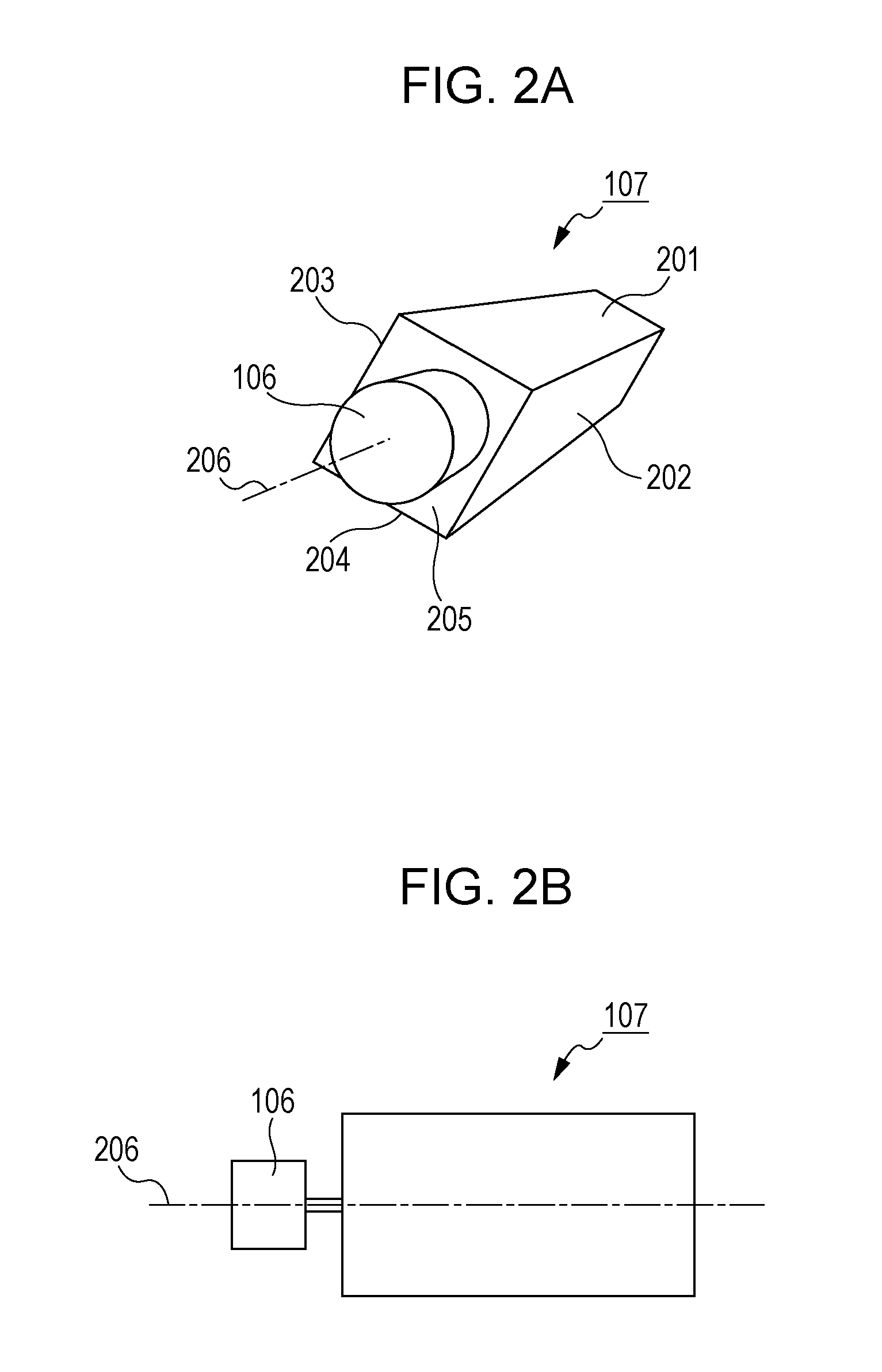
[0073]With the third embodiment, a configuration example wherein a linearly-driven actuator is used as the actuator for driving the specimen holder, rather than the rotational actuator configured of an ultrasonic motor as with the first embodiment, will be described with reference to FIGS. 8A and 8B. As shown in FIGS. 8A and 8B, multiple specimens 802, 803, and 804 are set in one row on the face of a specimen holder 801, with a linearly-driven actuator 805 being connected to the specimen holder 801. The linearly-driven actuator 805 moves the specimen holder 801 in the direction indicated by the arrow 806.

[0074]A shielding plate 809 having an observation window 808 is placed between the subject's eye 807 and the specimen holder 801, so that just one of the multiple specimens 802, 803, and 804 is presented to the subject. Also, illumination light is cast on the multiple specimens 802, 803, and 804 on the specimen holder 801 from an optical fiber illumination apparatus 810. The linearl...


PUM


Abstract

A visual stimulation presentation apparatus including: a specimen holding unit, made of a non-magnetic material, configured to hold multiple specimens of actual objects serving as specimens for presenting a subject with visual stimulation when running an apparatus to measure brain functions of the subject by detecting change in magnetic field; an actuator, made of a non-magnetic material, configured to selectively present multiple specimens of actual objects set in the specimen holding unit, by driving the specimen holding unit; and a control unit configured to control driving of the actuator; is used, whereby presenting time and presenting position can be precisely controlled and the delicate surface state of the objects with a high degree of sense of texture.

Description

BACKGROUND OF THE INVENTION[0001]1. Field of the Invention[0002]The present invention relates to a visual stimulation presenting apparatus used for presenting visual stimulation to a subject at the time of running an apparatus to perform brain function measurement of the subject by detecting change in magnetic field, and to a functional magnetic resonance imaging apparatus, a magnetoencephalograph apparatus, and a brain function measurement method.[0003]2. Description of the Related Art[0004]Apparatuses for measuring brain function by detecting change in magnetic field are used in various situations in medical practice. Magnetic resonance imaging (MRI) apparatuses and magnetoencephalograph (MEG) apparatuses are representative of apparatuses which measure brain function by detecting change in magnetic field. Of these, magnetic resonance imaging apparatuses are apparatuses which apply a magnetostatic field to the part of the subject to be measured, and further apply a particular high-...

Claims


Application Information

Patent Timeline
Patent Type & Authority Applications(United States)
IPC IPC(8): A61B5/05
CPCG01R33/4806
Inventor ICHIMURA, YOSHIKATSUKAWASHIMA, YASUHIRO
Owner CANON KK
Why Eureka
Try Eureka